Free NHA CET Practice Test: 50 Questions With Answers
Work through 50 original, unofficial NHA CET practice questions with answers and explanations for rhythm analysis, patient care, safety, and EKG technique.
Free NHA CET Practice Test
This free NHA CET practice test has 50 original questions for NHA's Certified EKG Technician exam, with answers and explanations. It follows the current test plan, is unofficial and is not a full-length exam; no sign-up is required.
Practice questions
Question 1 of 50
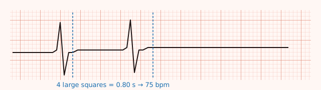
A rhythm strip is recorded at the standard paper speed of 25 mm per second. The rhythm is regular, and exactly four large squares separate consecutive R waves. What is the ventricular rate?
- A. 60 beats per minute
- B. 100 beats per minute
- C. 75 beats per minute
- D. 150 beats per minute
Reveal answer
C — 75 beats per minute.
Four large squares is 20 small squares, which is 20 mm of paper. At 25 mm per second, 20 mm takes 20 ÷ 25 = 0.8 seconds. One beat every 0.8 seconds is 60 ÷ 0.8 = 75 beats per minute.
The shortcut gets you there faster: at 25 mm/s, divide 300 by the number of large squares between R waves. 300 ÷ 4 = 75.
A would need five large squares, B three, and D two. Memorising the ladder — 300, 150, 100, 75, 60, 50 for one through six large squares — is worth learning.
Enlarge this educational schematic
Domain 3, Task A — calculate heart rate
Source: Utah ECG definitions (ECG Waves and Intervals; Bipolar and augmented limb leads). Arithmetic uses the stated inputs.
Question 2 of 50
A standard 10-second tracing shows 11 QRS complexes. The R-to-R intervals vary noticeably from beat to beat. What is the best way to report the ventricular rate?
- A. 66 beats per minute, from 11 complexes across 10 seconds
- B. 110 beats per minute, from 11 complexes multiplied by 10
- C. Divide 1500 by the shortest R-to-R interval in small squares
- D. Use the 300-150-100 sequence starting at the first R wave
Reveal answer
A — 66 beats per minute.
Ten seconds is one sixth of a minute, so 11 complexes across the strip works out to 11 × 6 = 66 beats per minute. Counting across the whole window averages the beat-to-beat variation, which is exactly what you want when the rhythm is irregular.
Use the duration actually printed on the recording; do not assume every displayed rhythm strip is ten seconds long.
B multiplies by 10, which is the rule for a six-second strip, not a ten-second one. C and D both measure one interval and treat it as the patient's rate. On an irregular rhythm that can be badly wrong — the fastest interval might read 100 while the slowest reads 55, and neither is the answer.
Domain 3, Task A — calculate heart rate
Source: Utah ECG definitions (ECG Waves and Intervals; Bipolar and augmented limb leads). Arithmetic uses the stated inputs.
Question 3 of 50
Your department uses 25 mm/s for routine resting recordings. You pick up a strip and the complexes look strangely spread out. Counting large squares the usual way, you calculate a rate of about 38 beats per minute — but the patient's radial pulse is about 75 and she feels fine. The machine was left set to 50 mm per second. What happened?
- A. Nothing is wrong. The patient is bradycardic and the pulse count is mistaken.
- B. The gain is doubled. Halve it and repeat.
- C. The paper speed is doubled, so anything measured as if it were 25 mm/s reads about twice its true duration. Reset to 25 mm/s and repeat.
- D. The high-frequency filter is switched on and is stretching the tracing.
Reveal answer
C — the paper speed is doubled.
The 300-divided-by-large-squares shortcut assumes 25 mm per second. At that speed a small square is 0.04 seconds and a large square is 0.20 seconds. Double the paper speed and the paper is the same but the time it represents is halved — a small square becomes 0.02 seconds.
Check the numbers. A true rate of 75 means an R-to-R of 0.8 seconds. At 50 mm/s that 0.8 seconds spans 40 mm, or 8 large squares. Apply 300 ÷ 8 and you get 37.5 — about half the real rate. Reset the machine, repeat, and if a non-standard speed was used deliberately, mark it on the tracing.
B confuses the two axes: gain changes height, speed changes width. A dismisses the mismatch instead of checking it. A manual pulse count is useful evidence, not an infallible measurement. D is invented — filters affect the signal, not the time scale.
Domain 3, Tasks A and C — heart rate and graph paper units
Source: QAS ECG acquisition procedure (PDF p. 3: preparation, position, chest-electrode table; p. 6: measurement units; p. 9: recording annotations). Arithmetic uses the stated inputs.
Question 4 of 50
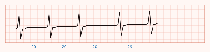
On a strip recorded at 25 mm/s, measuring consecutive R-to-R intervals, you get, in small squares: 20, 20, 20, 29, 20, 20, 20, 29. P waves are present and uniform. How should you describe this rhythm's regularity?
Enlarge this educational schematic
- A. Regular
- B. Regularity cannot be assessed without a full 12-lead
- C. Irregularly irregular — there is no pattern at all
- D. Regularly irregular — the long intervals repeat in a predictable pattern
Reveal answer
D — regularly irregular.
Three ways to describe regularity, and they're worth using precisely:
- Regular — R-to-R intervals essentially constant.
- Regularly irregular — the irregularity itself repeats. Grouped beating, like the pattern here, belongs in this bucket.
- Irregularly irregular — no repeating pattern anywhere.
Here, 20 small squares is 0.80 seconds and 29 is 1.16 seconds, and the long one arrives after every third short one. That repeating group is worth writing down, but the R-to-R pattern alone does not establish its cause. Naming the pattern accurately is yours.
A overlooks the recurring longer interval. C is the trap — spotting "not regular" and stopping there. B is wrong: regularity is measured on any adequate rhythm strip.
Domain 3, Task B — regularity
Source: How to measure the pulse (Procedure steps 4–6 and 8). Arithmetic uses the stated inputs.
Question 5 of 50
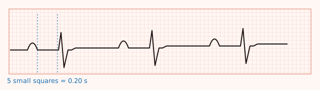
On an adult patient’s tracing recorded at 25 mm per second, the PR interval spans exactly five small squares in every beat, and every P wave is followed by a QRS. How should you record this?
Enlarge this educational schematic
- A. 0.05 seconds — abnormally short
- B. 0.20 seconds — first-degree AV block
- C. 0.50 seconds — markedly prolonged
- D. 0.20 seconds — at the upper limit of normal, not first-degree AV block
Reveal answer
D — 0.20 seconds, at the upper limit.
Five small squares × 0.04 seconds = 0.20 seconds.
The precision matters. First-degree AV block is defined as a PR interval greater than 0.20 seconds with all P waves conducting (University of Utah ECG Learning Center, Lesson 6). Exactly 0.20 is not greater than 0.20. It sits right at the boundary, and calling it first-degree AV block on that measurement alone would misapply the definition.
C reads each square as 0.10 seconds. A reads them as 0.01. Both are arithmetic slips that turn a normal tracing into an abnormal one, which is why fixing the square values in your head is worth more than memorising any rhythm name.
Domain 3, Task C — measure intervals
Source: Utah AV-block lesson (First-degree AV block; Type I and Type II second-degree AV block; 2:1 limitation). Arithmetic uses the stated inputs.
Question 6 of 50
At 25 mm per second, an adult patient’s QRS complex spans about three and a half small squares from its onset to its end. What duration do you record?
- A. 0.014 seconds — too narrow to be meaningful
- B. 0.14 seconds — wider than 0.12 seconds; report the measurement with the tracing
- C. 0.14 seconds — within normal limits, no comment needed
- D. 0.35 seconds — markedly prolonged
Reveal answer
B — 0.14 seconds.
3.5 × 0.04 = 0.14 seconds. That's wider than the roughly 0.12-second mark where conduction delay is usually described, so it's worth reporting rather than passing over.
Measure the interval, not just the sharpest visible part of the R wave. QRS duration runs from the beginning to the end of ventricular depolarization; a duration alone does not establish the cause of a widened complex.
C has the arithmetic right and the judgement wrong. A and D are decimal-place errors.
Domain 3, Tasks C and D — measure intervals and inspect waveforms
Sources: Utah ECG definitions (ECG Waves and Intervals; Bipolar and augmented limb leads); QAS ECG acquisition procedure (PDF p. 3: preparation, position, chest-electrode table; p. 6: measurement units; p. 9: recording annotations). Arithmetic uses the stated inputs.
Question 7 of 50
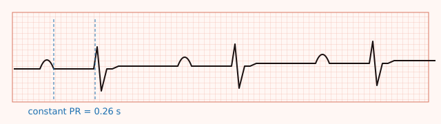
Rhythm strip. Lead II, 25 mm/s, standard calibration. Rate 68 and regular. A P wave precedes every QRS, and every P wave is followed by a QRS. The PR interval is constant at 0.26 seconds. QRS duration is 0.08 seconds.
Enlarge this educational schematic
- A. First-degree AV block
- B. Normal sinus rhythm
- C. Type I (Wenckebach) second-degree AV block
- D. Third-degree AV block
Reveal answer
A — first-degree AV block.
Two features, both required: a PR interval greater than 0.20 seconds, and every P wave conducting through to a QRS (Utah ECG Learning Center, Lesson 6). At 0.26 seconds with nothing dropped, that's what you have. The rate and the QRS width are both ordinary, which is typical.
B ignores the prolonged PR. C requires the PR interval to lengthen from beat to beat and then a P wave not to be followed by a QRS — neither happens here; the PR is constant and nothing is dropped. D would show the atria and ventricles running independently, with P waves marching through at their own rate and no fixed relationship to the QRS complexes.
"First-degree AV block" describes a conduction finding on a tracing. What it means for this patient is the provider's read.
Domain 3, Task E — identify arrhythmias
Source: Utah AV-block lesson (First-degree AV block; Type I and Type II second-degree AV block; 2:1 limitation).
Question 8 of 50
Rhythm strip. Lead II, 25 mm/s, standard calibration. P-to-P intervals are constant at 0.80 seconds. The PR intervals run 0.16, then 0.26, then 0.32 seconds; the next P wave has no QRS after it, and then the sequence starts over. QRS duration is 0.08 seconds throughout.
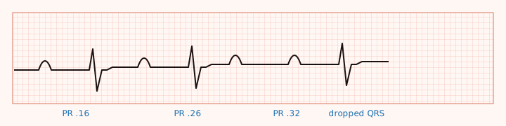
Enlarge this educational schematic
- A. First-degree AV block
- B. Type I (Wenckebach) second-degree AV block
- C. Type II (Mobitz) second-degree AV block
- D. Sinus rhythm with a premature beat
Reveal answer
B — Type I, or Wenckebach.
The signature is progressive PR lengthening ending in a P wave that doesn't conduct, then the cycle repeating. Notice the increments get smaller as it goes — 0.16 to 0.26 is a jump of 0.10, then 0.26 to 0.32 is only 0.06. That shrinking increment is part of the classic description, and it's why the pause after the dropped beat ends up shorter than two of the intervals before it. The QRS staying narrow fits too: Type I block usually sits in the AV node (Utah ECG Learning Center, Lesson 6).
C is the one to rule out carefully. In Type II the PR intervals stay constant before a P wave fails to conduct. Here they clearly lengthen. A has no dropped beats at all. D doesn't fit either — the P-to-P timing never varies, so nothing arrives early.
Domain 3, Task E — identify arrhythmias
Source: Utah AV-block lesson (First-degree AV block; Type I and Type II second-degree AV block; 2:1 limitation). Arithmetic uses the stated inputs.
Question 9 of 50
Rhythm strip. Lead II, 25 mm/s, standard calibration. P waves march out regularly at a P-to-P interval of 0.80 seconds. Every second P wave is followed by a QRS; the others are not. Every conducted beat has a PR interval of exactly 0.20 seconds. QRS duration is 0.08 seconds.
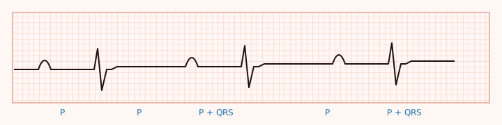
Enlarge this educational schematic
- A. 2:1 AV block — this strip does not contain two consecutive conducted beats, so Type I and Type II cannot be told apart from it
- B. Type I (Wenckebach) second-degree AV block, because beats are being dropped
- C. Type II (Mobitz) second-degree AV block, because the PR intervals are constant
- D. Third-degree AV block
Reveal answer
A — call it 2:1 AV block and stop there.
To call a block Type II you need to see two consecutive constant PR intervals before the dropped beat. In 2:1 conduction you never get two conducted beats in a row, so that comparison is impossible — and by the same logic you can't watch a PR interval lengthen either. The Utah ECG Learning Center says it plainly: with 2:1 AV block, "we can't be sure if it's type I or II" (Lesson 6).
C is the trap, and it's tempting: the PRs are constant. But those conducted beats are separated by nonconducted P waves. You cannot observe the PR trend across consecutive conducted beats in this 2:1 sequence. B invents lengthening that isn't there. D would show P waves and QRS complexes with no fixed relationship at all; here every conducted beat has the same PR, so they're clearly related.
Record what you measured — regular P waves, 2:1 conduction, PR 0.20 seconds on conducted beats, narrow QRS — and route it. Precision beats a confident wrong label.
Domain 3, Task E — identify arrhythmias
Source: Utah AV-block lesson (First-degree AV block; Type I and Type II second-degree AV block; 2:1 limitation).
Question 10 of 50
A patient with a documented demand pacemaker has a tracing showing her previously documented intrinsic wide-QRS rhythm at 70 per minute. Her device-clinic note states that pacing is inhibited when an intrinsic beat is sensed above the programmed lower rate. You can't see any pacing spikes anywhere on the page. A coworker says the device must have failed. What is the appropriate response?
- A. Agree, and tell the patient her pacemaker isn't working
- B. Note that a demand pacemaker may not deliver a stimulus when intrinsic beats are sensed, document what you actually observed, and route the tracing to the provider
- C. Turn the gain up until spikes appear
- D. Switch on the 40 Hz filter to sharpen the spikes
Reveal answer
B — absence of a visible spike does not establish device failure.
Demand pacemakers do not have to stimulate every beat. The sensing function can inhibit pacing when intrinsic beats occur above the programmed lower rate. That is a supported explanation for this patient’s recording; it is not proof that every aspect of the device is functioning normally.
A turns a single observation into an unsupported device-failure diagnosis. C changes the recorder’s gain but cannot make a pacemaker deliver a stimulus. D cannot establish device function by changing an ECG filter.
Document the actual recording and route it for review rather than telling the patient that her pacemaker has failed.
Domain 3, Task F — pacemaker activity
Source: AHA pacemaker explanation (How does a pacemaker work?).
Question 11 of 50
The machine prints an interpretation line reading "Anterior infarct, age undetermined." Reviewing your own work, you realise you placed V1 and V2 in the second intercostal space instead of the fourth. The patient has no symptoms. Your facility requires corrected recordings to be retained with the original and the acquisition error documented. What do you do?
- A. File the tracing — the computer read it, so it stands
- B. Delete the first tracing and say nothing
- C. Repeat the tracing with V1 and V2 correctly placed in the fourth intercostal space, and route both tracings to the provider with a note about the placement
- D. Edit the interpretation line to read "normal"
Reveal answer
C — repeat it correctly and be transparent about why.
V1 and V2 belong in the fourth intercostal spaces at the right and left sternal borders. Placing them in the second intercostal spaces changes the views being recorded. Correct the known acquisition error and label the repeat so the provider can interpret it in context.
The machine’s interpretation is not a reason to ignore a known placement error. Nor does finding that error prove the patient has no cardiac disease.
A treats a machine statement as a diagnosis. B hides a known error and violates the record-retention procedure given in the question. D substitutes an unsupported interpretation instead of correcting the acquisition and routing the tracing for review.
Reproducible placement makes the next comparison more useful too.
Domain 3, Tasks D and G — waveform characteristics and ischemia patterns
Sources: QAS ECG acquisition procedure (PDF p. 3: preparation, position, chest-electrode table; p. 6: measurement units; p. 9: recording annotations); NHA Candidate Handbook (Code of Ethics, printed p. 27; the role/record rule is supplied in the question).
Question 12 of 50
A telemetry alarm shows a wide-complex tachycardia at about 180 per minute. You go to the room. The patient is awake and talking, says she can feel her heart racing, and mentions she feels a little lightheaded. What is your immediate action?
- A. Reassure her it's probably artifact and re-check the strip later
- B. Leave the room to find the provider
- C. Print the strip and leave it for rounds
- D. Stay with her, activate your facility's escalation pathway immediately, and keep her on the monitor
Reveal answer
D — stay, escalate, keep monitoring.
You did the most important thing already by going to look at the patient. A sustained wide-complex tachycardia in a symptomatic patient is escalated now, not at the end of your list. Staying with her matters because her condition can change in the time it takes to walk down a corridor, and because you are the person watching the monitor.
A dismisses a finding without evidence. Being awake does not rule out a significant tachyarrhythmia; symptoms make prompt clinical assessment especially urgent. B leaves a symptomatic patient alone; use the call system. C treats a time-critical rhythm as paperwork.
Recognise, stay, escalate, document. Follow your training and the emergency-response protocol if her condition changes; do not withhold an indicated emergency response while waiting for an interpretation.
Domain 3, Task H — action on life-threatening arrhythmias
Sources: AHA adult advanced life support (Section 15, Wide-Complex Tachycardia, synopsis and recommendations); AHA adult BLS (Recognition of cardiac arrest and initiation of CPR; adult healthcare-professional algorithm).
Safety, compliance, and coordinated patient care (16 questions)
Question 13 of 50
A patient's employer calls the clinic. He says the patient needs a return-to-work clearance and asks you to confirm whether her EKG was normal. There is no written authorization, and the records team confirms that no legally permitted exception applies to this request. Your facility routes employer requests through that team. What should you do?
- A. Decline, and direct the request to the process your facility uses for records requests — this disclosure needs the patient's written authorization
- B. Say only whether it was normal or abnormal, with no detail
- C. Confirm it, since the employer is paying for the visit
- D. Ask the patient over the phone for permission, then tell him
Reveal answer
A — this one needs written authorization, and it isn't your call to process.
The Privacy Rule permits use and disclosure without authorization for treatment, payment and health care operations. This employer request has no applicable permission or exception in the facts given. HHS names this situation specifically: disclosing the results of a pre-employment physical or lab test to an employer is an example of a disclosure that requires the individual's written authorization (HHS, Summary of the HIPAA Privacy Rule, "Authorized Uses and Disclosures").
C confuses paying a bill with a right to the clinical result. B is still a disclosure — "normal" is protected health information about an identified person. D gets the form wrong: the authorization needed for this request must be written and specific. It also bypasses the records process stated in the question.
Domain 1, Task A — HIPAA
Source: HHS Privacy Rule summary (Authorized Uses and Disclosures; Public Interest and Benefit Activities (exceptions excluded by the stem)).
Question 14 of 50
The office of the cardiologist your patient was referred to calls and asks for a copy of the tracing you recorded this morning so she can review it before the appointment. What is correct?
- A. Verify the requester’s identity and authority, then release the requested tracing through your facility’s normal process for treatment-related requests
- B. Refuse until the patient signs a written authorization
- C. Release it only after removing the patient's name and date of birth
- D. Explain that the minimum necessary rule blocks any release outside your facility
Reveal answer
A — this is a treatment disclosure, and it's permitted.
Treatment includes consultation between providers about a patient and referral of a patient from one provider to another. Those disclosures are permitted without the patient's authorization. There's a second point worth knowing: the minimum necessary standard does not apply to a disclosure to, or a request by, a health care provider for treatment (HHS, Summary of the HIPAA Privacy Rule, "Limiting Uses and Disclosures to the Minimum Necessary").
Put Questions 13 and 14 side by side. Same tracing, same technician, opposite answers — because the purpose of the request is what decides.
B over-restricts and delays care. C strips the identifiers the cardiologist needs. D misstates the rule in exactly the direction people get it wrong.
Domain 1, Task A — HIPAA
Sources: HHS Privacy Rule summary (Permitted Uses and Disclosures—Treatment; Limiting Uses and Disclosures to the Minimum Necessary); 45 CFR 164.514(h) (Paragraph (h)(1), verification requirements).
Question 15 of 50
You've just removed your gloves after finishing an EKG. Your next patient is in a room across the hall. What should you do right now?
- A. Nothing — the gloves protected your hands
- B. Perform hand hygiene now, immediately after removing the gloves
- C. Wash with soap and water only if you can see soiling on your hands
- D. Wait and perform hand hygiene just before touching the next patient
Reveal answer
B — hand hygiene is indicated immediately after glove removal.
CDC lists it as one of the clinical indications for hand hygiene, alongside immediately before touching a patient, after touching a patient or the patient's immediate environment, and after contact with blood, body fluids or contaminated surfaces (CDC, Core Infection Prevention and Control Practices, section 5a). Gloves are a barrier, not a substitute.
D is the interesting wrong answer, because it names a real indication — you do owe hand hygiene before touching the next patient. It just doesn't cancel the one you owe now. Two separate moments.
C mixes up the two rules. Visible soiling determines the product (soap and water rather than alcohol-based rub); it doesn't determine whether hand hygiene happens.
Domain 1, Task B — infection control
Source: CDC core infection-prevention practices (Section 5a, indications immediately after glove removal).
Question 16 of 50
You finish a 12-lead. The disposable electrodes are peeled off and discarded, and you coil the reusable lead wires and set them on the machine's supply shelf next to an unopened pack of fresh electrodes. What is wrong with this?
- A. Nothing — the lead wires clip onto electrodes and never touch skin
- B. The wires should have been discarded along with the electrodes
- C. The wires should have been reprocessed according to the manufacturer's instructions and kept away from clean supplies
- D. It only matters if the next patient is on transmission-based precautions
Reveal answer
C — reprocess them, and keep clean and soiled separate.
Reusable medical equipment is cleaned and reprocessed before use on another patient or when soiled, following the manufacturer's instructions for that specific device, and clean equipment is kept separated from soiled equipment to prevent cross-contamination (CDC, Core Infection Prevention and Control Practices, section 5f). Lead wires and clips can become contaminated through contact with the patient, gown, bedding or your gloves, even when an electrode sits between a clip and the skin.
A assumes the reusable wires cannot become contaminated. B wastes reusable equipment; the answer is reprocessing, not the bin. D inverts standard precautions, which apply to every patient regardless of known infection status.
Why the manufacturer's instructions rather than a rule of thumb? Because contact time, dilution and material compatibility differ by product, and an incompatible agent may damage equipment or fail to provide the required disinfection.
Domain 1, Task B — infection control
Source: CDC core infection-prevention practices (Section 5f, reprocessing and separating clean/soiled equipment).
Question 17 of 50
Your job description permits you to document measured findings but reserves the final diagnostic interpretation for an authorised clinician. A nurse who is short-staffed asks you to enter that final interpretation in the chart so the note is complete. The tracing looks like ordinary sinus rhythm to you. What do you do?
- A. Enter "normal sinus rhythm" — that's what it is
- B. Enter "abnormal" so the chart flags it for review
- C. Leave the EKG out of the record until the provider dictates a reading
- D. Enter the objective findings you obtained and route the tracing for provider interpretation
Reveal answer
D — document what you measured, not what it means.
An EKG technician records the tracing and documents observable, measurable findings: rate, regularity, intervals, artifact, position and lead modifications, and the patient's condition during the recording. Entering the final diagnostic interpretation is outside the role described in this question, and being asked nicely by a busy colleague doesn't extend it.
A is the natural mistake because the reading is probably right. Being right isn't the issue — a final diagnostic statement entered under your name would exceed the authority specified in your job description, and once it's in the chart, someone will rely on it.
C overcorrects. The tracing belongs in the record now; delaying it can bury a time-sensitive finding.
B substitutes an unsupported label for the measured findings the question permits you to document. A misleading flag is not a substitute for review.
Domain 1, Task C — scope of practice
Source: NHA Candidate Handbook (Code of Ethics, printed p. 27; the role/record rule is supplied in the question).
Question 18 of 50
After you finish, a patient asks you to take the tracing out of her record. She says it looks bad and she doesn't want it seen. How do you respond?
- A. Delete it and record a fresh one
- B. Mark the first tracing "invalid" yourself
- C. Repeat the tracing and file only the better-looking one
- D. Explain that you cannot delete it on this request, and tell her how to discuss it with her provider or request an amendment through medical records
Reveal answer
D — you can't remove it, and she has a route that isn't you.
A completed diagnostic recording is part of the medical record. A patient’s concern does not authorise you to delete or invalidate it outside the facility’s correction process. Doing so could hide a finding that needs attention.
The part that makes this a good answer rather than a brush-off is the second half. Patients do have rights here: they can ask a provider about the finding and request an amendment if they believe the record is inaccurate or incomplete. An amendment request is not an automatic right to erase an accurate result. Point her at the real process instead of leaving her with "no."
A, C and B are three versions of the same act — deciding on your own which version of the truth goes in the chart.
Domain 1, Task C — scope and ethical standards
Sources: HHS Privacy Rule summary (Individual Rights—Amendment); NHA Candidate Handbook (Code of Ethics, printed p. 27; the role/record rule is supplied in the question).
Question 19 of 50
Your patient is deaf and reads lips. You're wearing a surgical mask, and he's clearly not following your instructions. What is the appropriate approach?
- A. Speak more loudly through the mask
- B. Have his adult daughter in the waiting room relay everything
- C. Ask him how he communicates best and use that method — for example, written instructions
- D. Skip the explanation and just position him
Reveal answer
C — ask him, then use what works.
The patient is the authority on how he communicates. Some people will want written instructions, some a clear-panel mask if your facility stocks them, some a qualified interpreter arranged through your facility. Ask which aid or service works for him, and arrange effective communication for the complexity of the discussion.
A is the reflex answer and it doesn't help — volume isn't the barrier; the mask covering your lips is. B assumes his daughter should interpret without asking him or arranging an appropriate aid. A family member is not the default substitute for effective communication; the ADA permits reliance on an accompanying adult only in limited circumstances. D removes his ability to consent to and cooperate with the procedure, which also makes for a worse tracing.
Domain 1, Task D — communication
Source: DOJ effective-communication guidance (Choosing effective aids; Use of Accompanying Adults or Children as Interpreters).
Question 20 of 50
An order in your queue reads "EKG" with no further detail, and you can't tell whether the provider wants a resting 12-lead or a rhythm strip. No standing protocol resolves the ambiguity. What should you do?
- A. Do a 12-lead — it contains more information
- B. Do both to cover the possibilities
- C. Ask the patient which one her doctor meant
- D. Clarify the order with the ordering provider or the nurse before you start
Reveal answer
D — clarify before you acquire.
Clarify an ambiguous order rather than guessing and risking a repeat. Clarifying is a routine part of working inside a care team, not an admission that you don't know your job.
A sounds efficient and is a decision about what test the patient receives — which is the provider's. B adds a test without resolving which test was requested. C asks the patient to be responsible for the accuracy of a clinical order.
Domain 1, Task D — coordinated care
Source: NHA Candidate Handbook (Code of Ethics, printed p. 27; the role/record rule is supplied in the question).
Question 21 of 50
Before a routine resting EKG you check the patient's radial pulse and find it irregular. You cannot get a reliable count in 15 seconds. What do you do?
- A. Count for a full 60 seconds and document the irregularity along with the rate
- B. Count for 15 seconds and multiply by four
- C. Skip the manual pulse — the EKG will produce a rate
- D. Take the reading from the pulse oximeter instead
Reveal answer
A — full minute, and say that it was irregular.
A 15-second count multiplied by four assumes the next 45 seconds look like the first 15. On an irregular pulse they don't, and small counting windows magnify the error — one extra or missing beat in 15 seconds moves the reported rate by four. Counting the full minute averages the variation.
Documenting the irregularity matters as much as the number. The person reading the tracing later may see a shorter recording window; you were at the bedside for a minute. That observation is genuinely yours to contribute.
B is the standard shortcut applied where its assumption fails. C and D substitute a device reading for a clinical observation, and neither one tells the provider what your fingers told you.
Domain 1, Task E — vital signs
Source: How to measure the pulse (Procedure steps 4–6 and 8). Arithmetic uses the stated inputs.
Question 22 of 50
While you’re explaining an exercise stress test, the patient asks, “Can I ask to stop if I need to, even before I reach the target heart rate?” What is the accurate thing to tell her?
- A. “Only if the provider gives permission after you request it.”
- B. "We keep going until you hit your target heart rate, no matter what."
- C. “Yes. Tell us immediately if you need to stop or develop symptoms such as chest pain or dizziness. The team will help you stop safely, and may also stop the test for clinical reasons.”
- D. "Try to push through — stopping early makes the test useless."
Reveal answer
C — accurate, and it gives her something to do.
Three true things fit together: report symptoms immediately, you can stop if you need to, and the clinical team can also end the test for safety. She leaves the conversation knowing her role and knowing she isn't trapped on the treadmill.
A makes stopping depend on permission the patient does not need to withdraw from the test. B is both wrong and unsafe — a target heart rate is not a reason to keep going through symptoms. D pressures a patient to suppress exactly the information the test depends on her reporting.
The EKG technician's job here is instruction and monitoring, not setting or overriding the stopping criteria.
Domain 1, Task F — stress testing instruction
Source: AHA stress-test guidance (PDF p. 1: preparation and alternative stress; p. 2: stopping and recovery).
Question 23 of 50
A patient scheduled for a treadmill stress test tells you at check-in that he can only walk a few steps with a frame because of a recent hip fracture. What is the appropriate action?
- A. Flag the mobility limitation to the ordering provider before the appointment proceeds, since a medication-based (pharmacologic) stress test may be ordered instead
- B. Have him walk as far as he can and stop there
- C. Cancel the test and tell him to rebook
- D. Run the standard protocol at the lowest treadmill speed
Reveal answer
A — flag it; a different type of stress test may be the answer.
Exercise isn't the only way to stress the heart. When a patient can't exercise adequately, a pharmacologic stress test — where a medication produces the stress instead of a treadmill — is one of the options a provider can order. Recognising that the ordered test may not fit the patient in front of you, and saying so before anyone steps on a treadmill, is squarely part of the technician's role.
B puts an unsteady patient on a moving belt without addressing the limitation. D changes the protocol without an authorised plan. C sends him away before the clinical team can decide the appropriate next step.
Choosing the alternative protocol is the provider's decision. Surfacing the problem in time is yours.
Domain 1, Task F — stress testing
Source: AHA stress-test guidance (PDF p. 1: preparation and alternative stress; p. 2: stopping and recovery).
Question 24 of 50
A patient is prescribed a 30-day, patient-activated external loop event recorder. He asks how it differs from the 24-hour Holter monitor he wore last year. What is accurate?
- A. They're the same device with different brand names
- B. An event monitor is worn only while the patient is asleep
- C. An event monitor records continuously and a Holter records only when triggered
- D. His Holter stored a continuous recording for its prescribed period; this loop recorder samples continuously but saves selected segments around activation, following this device’s instructions
Reveal answer
D.
The two answer different questions. A Holter stores a continuous ECG over the prescribed recording period. The external loop recorder in this question samples continuously into a short rolling memory and saves a segment when activated, including activity before the button press. That lets the clinician examine the ECG around an episode.
That difference changes your instructions. A Holter patient needs to wear it and log what happened and when. This event-recorder patient also needs to know how to activate and transmit a recording as instructed.
C reverses the devices’ roles. A collapses a real distinction. B is invented, and would defeat the point of monitoring symptoms during ordinary activities.
Not all event monitors work this way: some detect events automatically, some are handheld, and some patch monitors record continuously. Always teach from the instructions for the device in your hand — wear time, water exposure and triggering are device-specific and facility-specific.
Domain 1, Task G — ambulatory monitoring
Sources: AHA cardiac event recorder (Types of cardiac event recorders; What should I expect?); AHA Holter monitor (What is a Holter monitor?; What should I expect?).
Question 25 of 50
You've finished explaining the event monitor. You ask, "Any questions?" and the patient says no. What should you do next?
- A. Accept that as confirmation and move on
- B. Hand him the manufacturer's pamphlet and let him read it at home
- C. Repeat the instructions once more, more slowly
- D. Ask him to describe in his own words what he'll do the next time he feels his heart flutter
Reveal answer
D — have him tell you the plan back.
The CET test plan doesn't just ask you to instruct patients on ambulatory monitors; it asks you to verify their understanding. "No questions" doesn't do that. People say it when they're embarrassed, rushed, or don't yet know enough to know what to ask.
Asking him to walk through what he'll actually do turns a yes/no into something you can check — and if a step is missing, you find out while he's still in front of you.
C repeats the message without checking whether it landed. B is a fine supplement and a poor substitute; you learn nothing about whether he can use the device. A is the one that feels like a complete interaction and isn't.
Domain 1, Task G — verifying understanding
Source: AHRQ teach-back guidance (Overview; Try the teach-back method; clarify and check again).
Question 26 of 50
You've completed a tracing and go to attach it to the chart. The EHR shows two patients with the same last name and very similar first names, both seen this morning. Your facility requires full name and date of birth to match the patient, order and destination record. What do you do?
- A. Choose the record whose appointment time matches yours
- B. Attach it to both records so nothing is missed
- C. Verify two patient identifiers against the patient and the order before attaching the tracing
- D. Save it to your desktop and sort it out later
Reveal answer
C — two identifiers, checked against the patient and the order.
Use the two identifiers required in this question: full name and date of birth. Match them across the patient, order, tracing and destination record before attaching the file. Looking twice at the same ambiguous screen is not an independent identity check.
A uses one weak identifier, and appointment times get moved. B puts one patient's tracing in another patient's record — a documented error affecting two people. D postpones the identification problem and creates an unnecessary separate copy of protected information instead of following the approved filing process.
Domain 1, Task H — electronic health record
Sources: HHS Privacy Rule summary (Permitted Uses and Disclosures; Authorized Uses and Disclosures; Minimum Necessary; Amendment; Administrative Requirements); NHA Candidate Handbook (Code of Ethics, printed p. 27; the role/record rule is supplied in the question).
Question 27 of 50
Halfway through a resting 12-lead, an adult patient who is already lying supine becomes pale and clammy and says she feels about to faint. She remains responsive and is breathing normally, with no injury or pregnancy-related concern. What do you do first?
- A. Finish the tracing quickly so you have a record of the episode
- B. Sit her upright so she can catch her breath
- C. Stop, keep her lying down, stay with her, and call for help using the room's call system
- D. Leave the room to find the nurse
Reveal answer
C — stop, keep her flat, stay, call from where you are.
New pallor, clamminess and near-fainting in a patient who is already lying down are a reason to stop and get help, not to assume a harmless cause. Two parts of the answer matter equally. Keeping her supine is protective if she's about to faint. Staying with her is protective because she is now someone who could lose consciousness with nobody watching.
D is the one that catches people, because going to get help feels like getting help. Use the call system; that's what it's for.
A prioritises a document over a patient. B moves a lightheaded patient upright, the wrong direction if she's about to pass out.
You are not diagnosing what caused this. You're stopping, protecting, summoning and staying.
Domain 1, Task I — cardiopulmonary compromise
Source: AHA syncope guidance (Vasovagal warning symptoms and assessment; higher-risk features including episodes while supine).
Question 28 of 50
You are a BLS-trained healthcare responder. The scene is safe, and you find an adult unresponsive in a treatment room. You've called for help and someone has gone for the AED. The patient is not breathing normally — only occasional gasps — and you cannot feel a definite pulse within 10 seconds. What is your next action?
- A. Check the telemetry monitor to see what the rhythm is
- B. Begin chest compressions
- C. Give two rescue breaths first, then reassess
- D. Wait for the AED to arrive before touching the patient
Reveal answer
B — start compressions.
Occasional gasping is not normal breathing. An unresponsive adult who isn't breathing normally, with no definite pulse found within 10 seconds, is treated as cardiac arrest, and early high-quality chest compressions with prompt defibrillation are the interventions most associated with survival (2025 AHA Guidelines for CPR and ECC, Part 7: Adult Basic Life Support).
A is the trap for people who work around monitors all day. The patient's condition, not the waveform, tells you what to do. D delays compressions for equipment that's already on its way — compressions start now, with interruptions kept brief for AED analysis and shock delivery as prompted. C reverses the sequence.
This question is about recognising arrest and starting. It isn't a substitute for BLS certification, and the CET test plan lists CPR and basic life support as core knowledge that can appear anywhere on the exam — worth knowing cold for that reason alone.
Domain 1, Task I — basic life support
Source: AHA adult BLS (Recognition of cardiac arrest; initiation of resuscitation; healthcare-professional algorithm).
EKG acquisition (22 questions)
Question 29 of 50
You open the electrode pouch and find it was left unsealed overnight. The gel on the electrodes feels tacky and dry at the edges. What should you do?
- A. Discard them and open a fresh, sealed pack
- B. Add a drop of conductive gel to each one and use them
- C. Use them — the adhesive still works
- D. Use them on the limb leads only, where contact matters less
Reveal answer
A — open a fresh pack.
Dried gel can cause a noisy or unusable tracing. Conduction between skin and electrode depends on that gel being intact.
B improvises a repair instead of using electrodes with intact gel as intended. C confuses adhesion with conduction; an electrode can stick perfectly and still record badly. D invents a hierarchy that doesn't exist: limb-electrode contact matters too, and one poor limb connection can affect several derived leads.
Domain 2, Task A — maintain equipment
Sources: QAS ECG acquisition procedure (PDF p. 3: preparation, position, chest-electrode table; p. 6: measurement units; p. 9: recording annotations); QAS cardiac-monitoring procedure (PDF p. 2, artifact causes); Bioelectromagnetism, chapter 15 (Sections 15.1, 15.3–15.7; equations 15.1–15.7).
Question 30 of 50
A colleague wipes down the EKG machine's screen and lead cables with a disinfectant wipe that isn't listed in the machine's cleaning instructions. Why does this matter?
- A. It doesn't — any hospital-grade disinfectant is interchangeable
- B. Products differ in contact time, dilution and material compatibility, so the wrong one may fail to disinfect and may damage the cables
- C. Only the electrodes need disinfection
- D. Cables should be cleaned with water only
Reveal answer
B.
CDC's core practices are specific on this point twice over: follow manufacturers' instructions for the proper use of cleaning and disinfecting products, including dilution, contact time, material compatibility, storage and shelf life; and consult and adhere to manufacturers' reprocessing instructions for reusable equipment (CDC, Core Infection Prevention and Control Practices, sections 5b and 5f).
Material compatibility matters even when a surface looks clean: an incompatible product can damage the equipment.
A treats "disinfectant" as one product. C ignores everything the patient and your gloves touched. D doesn't disinfect anything.
Domain 2, Task A — maintain equipment
Source: CDC core infection-prevention practices (Sections 5b and 5f, manufacturer instructions and material compatibility).
Question 31 of 50
Your department's standard calibration produces a calibration mark 10 mm tall for 1 millivolt. On the tracing you just printed, the calibration mark measures 5 mm, and nothing is noted on the page. What is the consequence?
- A. Waves print at half the height they would at 10 mm/mV; a reader who assumes standard gain will underestimate voltage
- B. None — calibration only affects the printed appearance
- C. All intervals will be measured as twice their true duration
- D. The heart rate calculation will be wrong
Reveal answer
A — half the standard printed height.
Gain sets the vertical scale: how many millimetres of paper represent one millivolt of signal. Halve it and every wave prints at half the height it would have at the original gain; the heart’s signal has not halved. That's a legitimate thing to do when complexes are so tall they overlap the channels above and below — but only if you mark the change on the tracing, so the scale is unambiguous. The calibration pulse itself is also a scale reference — here, a 1-mV pulse printed at 5 mm shows 5 mm/mV.
C and D both belong to the horizontal axis, which is paper speed. Gain doesn't touch timing, so the rate is unaffected.
Two habits follow from this. Look at the calibration mark before you read anything off a tracing. And if you change speed or gain deliberately, write it on the page.
Domain 2, Task B — verify machine settings
Source: QAS ECG acquisition procedure (PDF p. 3: preparation, position, chest-electrode table; p. 6: measurement units; p. 9: recording annotations). Arithmetic uses the stated inputs.
Question 32 of 50
Which describes the correct relationship between paper speed and gain at standard settings?
- A. Both settings affect amplitude only
- B. Paper speed sets amplitude; gain sets time
- C. Paper speed sets the horizontal (time) scale; gain sets the vertical (amplitude) scale
- D. Both settings affect time only
Reveal answer
C — speed is sideways, gain is up and down.
Increasing the paper speed is not a way to fix tall complexes, and halving the gain is not a way to fix long intervals.
Anchor it to the paper. At the standard 25 mm per second, one small square is 0.04 seconds and one large square is 0.20 seconds — that’s speed. Gain governs the other axis: how tall one millivolt prints. The page layout is a separate display choice, not something the paper speed dictates.
Change speed and the tracing stretches or compresses sideways. Change gain and it grows or shrinks vertically. Nothing else moves.
A and D assign both controls to one axis. B reverses the axes.
Domain 2, Task B — verify machine settings
Source: QAS ECG acquisition procedure (PDF p. 3: preparation, position, chest-electrode table; p. 6: measurement units; p. 9: recording annotations). Arithmetic uses the stated inputs.
Question 33 of 50
For this recording, the device instructions and your approved diagnostic protocol specify a high-frequency cutoff of 150 Hz. A colleague switches it to 40 Hz — labelled “muscle filter” — to smooth a shaky tracing. The patient is stable and visibly tensing her arms. What should you do?
- A. Restore the specified diagnostic setting, help the patient relax and support her arms, then repeat the recording
- B. Keep the 40 Hz setting because a smoother tracing is always a better diagnostic recording
- C. Keep the changed setting; writing “40 Hz” on the tracing overrides the specified protocol
- D. Ignore the setting change because high-frequency filtering affects only baseline drift
Reveal answer
A — fix the cause, and use the specified recording settings.
The question supplies the setting required for this device and protocol. A smoother-looking trace is not a reason to substitute a different mode. Here there is also a visible source to address: sustained muscle tension can introduce artifact.
B ignores the diagnostic setting stated in the question. C documents a change but does not make that change appropriate. D assumes filtering is harmless without checking the device instructions.
The real fix is upstream — relax and support the patient, re-prep the skin if needed, and check contact. Filter settings are device- and purpose-specific; the numbers in this scenario are not a universal NHA machine-setting rule.
Domain 2, Tasks B and G — machine settings and artifact
Source: QAS cardiac-monitoring procedure (PDF p. 2, artifact causes).
Question 34 of 50
Your patient has applied a thick body lotion across her chest this morning. What is the correct preparation?
- A. Apply extra conductive gel over the lotion
- B. Move the chest electrodes to lotion-free skin
- C. Tape the electrodes down firmly over the lotion
- D. Clean the electrode sites and let the skin dry completely before applying electrodes
Reveal answer
D — clean it off, let it dry.
Clean and dry the correct electrode sites before applying electrodes. Lotion is both an insulating layer and an adhesion problem, so it has to come off the sites.
B is the trap, and it's the one that does real damage. A precordial electrode's position is the measurement — move V3 somewhere convenient and you've changed what that lead records, without giving the reader the intended view. Prepare the correct site; never relocate a chest electrode for convenience.
A and C both try to bridge the barrier instead of removing it, and neither restores clean contact.
Domain 2, Task C — prepare the skin
Sources: QAS ECG acquisition procedure (PDF p. 3: preparation, position, chest-electrode table; p. 6: measurement units; p. 9: recording annotations); AHA cardiac event recorder (Types of cardiac event recorders; What should I expect?).
Question 35 of 50
The limb-lead baselines are noisy, and you notice the intact, non-fragile skin at the electrode sites is dry and visibly flaking. The electrode instructions and facility protocol permit gentle surface abrasion when needed. What's the appropriate step?
- A. Increase the gain so the complexes stand out from the noise
- B. Print it and note "poor skin condition"
- C. Use larger electrodes to cover more surface
- D. Clean the sites and gently abrade the skin according to your facility's protocol, then apply fresh electrodes
Reveal answer
D — clean and gently abrade.
Loose surface skin sits between the electrode and the tissue you're trying to record from. Follow the preparation instructions given in the question, then reassess the recording. Do not abrade damaged or fragile skin. "Gentle" is doing work in that sentence — you're removing loose surface cells, not scrubbing.
A amplifies noise and signal equally, so the ratio doesn't improve. C doesn't help if the contact surface is the problem rather than its size. B documents a problem you could have solved and hands the reader a degraded tracing.
Domain 2, Task C — prepare the skin
Sources: AHA cardiac event recorder (What should I expect?—preparing skin; device-specific preparation supplied in the stem); QAS ECG acquisition procedure (PDF p. 3: preparation, position, chest-electrode table; p. 6: measurement units; p. 9: recording annotations).
Question 36 of 50
A stable patient with severe COPD reports her usual difficulty lying flat and asks to stay propped up. You record a good-quality tracing with her semi-upright. What must you do?
- A. Note the non-standard position on the tracing so it is not silently compared as if recorded in the usual supine position
- B. Nothing further — the tracing is diagnostic
- C. Repeat it supine as soon as she can tolerate it
- D. Record it as a rhythm strip rather than a 12-lead
Reveal answer
A — record it in the position she can tolerate, and say so on the page.
Record the position actually used. A semi-recumbent position can accommodate a patient who cannot comfortably lie flat, and the record should make that difference clear to the reader. If breathlessness is new or worsening, escalate it rather than treating it only as a positioning problem.
B hands the reader a tracing that looks standard and isn't. C does not replace the required documentation and assumes another recording is necessary; follow the clinical team’s directions rather than deciding that from position alone. D downgrades the test the provider ordered.
Same principle throughout acquisition: when you deviate from standard for a good reason, deviate and document. What you never do is deviate silently.
Domain 2, Task D — position the patient
Sources: QAS ECG acquisition procedure (PDF p. 3: preparation, position, chest-electrode table; p. 6: measurement units; p. 9: recording annotations); NHA Candidate Handbook (Code of Ethics, printed p. 27; the role/record rule is supplied in the question).
Question 37 of 50
The limb leads show a jittery, irregular baseline. The patient is supine with her arms crossed tightly over her chest, gripping her own elbows. What should you do first?
- A. Ask her to relax her arms at her sides, supported by the table, then repeat
- B. Apply the 40 Hz filter and reprint
- C. Increase the gain
- D. Move the limb electrodes onto the torso
Reveal answer
A — relax and support the limbs.
Tensed muscle produces somatic artifact, and gripping her own elbows means both arms are working continuously. Supporting the limbs so no muscle group is holding a position removes the source. Address that source and check the repeat instead of only changing the display.
B changes filtering before addressing an obvious source and checking the required diagnostic settings. C amplifies the artifact too. D changes the electrode locations rather than first correcting the arm tension; a modified placement must be deliberate and documented.
Domain 2, Tasks D and G — positioning and artifact
Sources: QAS ECG acquisition procedure (PDF p. 3: preparation, position, chest-electrode table; p. 6: measurement units; p. 9: recording annotations); QAS cardiac-monitoring procedure (PDF p. 2, artifact causes); Bioelectromagnetism, chapter 15 (Sections 15.1, 15.3–15.7; equations 15.1–15.7).
Question 38 of 50
You have correctly located V4 and V6, but cannot confidently identify the anterior axillary line on this patient. Where should V5 go?
- A. In the fifth intercostal space, wherever the anterior axillary line seems to fall
- B. Midway between V4 and V6, in the horizontal plane of V4
- C. Directly below the nipple
- D. One intercostal space below V4
Reveal answer
B — midway between V4 and V6.
Use the midpoint between V4 and V6, keeping V5 in the same horizontal plane. The acquisition procedure linked below lists V5 midway between those two positions. Do not trace an intercostal space downward around the chest instead.
D follows the intercostal space downward, instead of keeping the electrode in the horizontal plane of V4. C uses a landmark that moves between patients and isn't a standard reference. A keeps estimating the line you already said you couldn't find.
Reproducibility is the whole point. Consistent precordial placement makes serial comparison more useful.
Domain 2, Task E — apply electrodes
Source: QAS ECG acquisition procedure (PDF p. 3, chest-electrode table: V5 midway between V4 and V6).
Question 39 of 50
A tracing shows lead I inverted, leads II and III appearing swapped, and aVR and aVL appearing swapped. Lead aVF looks unchanged, and all six chest leads look normal. What is the most likely cause?
- A. The right arm and right leg electrodes are reversed
- B. The left arm and right arm electrodes are reversed
- C. The patient has dextrocardia
- D. V1 and V2 are misplaced
Reveal answer
B — left arm and right arm reversed.
This has a fingerprint, and it's worth memorising because the pattern follows from the lead connections. Swapping the two arm electrodes inverts lead I, swaps leads II and III, swaps aVR and aVL, and leaves aVF untouched. The chest leads are unaffected because the reference used to derive them isn't disturbed (12-Lead ECG System, sections 15.1, 15.3 and 15.4; the reversal pattern follows by exchanging RA and LA in the lead relationships).
C is the answer worth knowing how to exclude, since dextrocardia can look similar in the limb leads. Check the actual cable connections rather than diagnosing dextrocardia from an inverted lead I. The complete swap pattern and preserved chest-lead recording described here point to the arm connections.
A produces a different picture — see the next question. D would change the chest leads, which look normal here.
Fix it and repeat. A reversal can create false-positive or false-negative signs of ischemia.
Domain 2, Task E — apply electrodes
Sources: Bioelectromagnetism, chapter 15 (Sections 15.1, 15.3–15.7; equations 15.1–15.7); Utah ECG definitions (ECG Waves and Intervals; Bipolar and augmented limb leads). The connection effects follow from the linked lead relationships.
Question 40 of 50
An awake patient has a palpable pulse. Lead II is nearly flat, while the other leads still show QRS complexes, although some shapes look different from the previous recording. Which connection error should you check among these options?
- A. Whether the right arm and right leg cables are reversed
- B. The V2 electrode
- C. The paper speed
- D. Whether the patient has asystole
Reveal answer
A — right arm and right leg reversed.
Lead II normally records the potential difference between the left leg and the right arm. Swap the right arm and right leg cables and lead II ends up recording the difference between the two legs, which is nearly zero — so it prints as a near-flat line while other leads can still show complexes. Their shapes need not remain normal. This pattern is a reason to inspect the connections, not proof of a particular reversal.
D does not fit the awake patient with a palpable pulse and QRS complexes in other simultaneous leads. A flat channel can be a recording problem; it does not by itself diagnose asystole. Always assess the patient as well as the tracing.
B would affect a chest lead, not lead II. C changes the time scale, not the amplitude.
Domain 2, Task E — apply electrodes
Sources: Utah ECG definitions (ECG Waves and Intervals; Bipolar and augmented limb leads); Bioelectromagnetism, chapter 15 (Sections 15.1, 15.3–15.7; equations 15.1–15.7); AHA adult BLS (Recognition of cardiac arrest and initiation of CPR; adult healthcare-professional algorithm). The connection effects follow from the linked lead relationships.
Question 41 of 50
A provider orders a right-sided V4R recording. Where does V4R go, and how do you label the tracing?
- A. At standard V4 on the left chest, recorded twice and labelled “repeat”
- B. On the patient's back, in the horizontal plane of V6
- C. At the fifth intercostal space on the right midclavicular line, with the recording clearly labelled V4R
- D. In the second intercostal space bilaterally
Reveal answer
C — V4’s right-sided counterpart, clearly labelled.
V4R is the right-sided counterpart of standard V4: the fifth intercostal space on the right midclavicular line. Follow the ordered lead set and label each actual position; do not assume that every facility uses the same repeat-recording sequence.
Labelling is not a nicety. An unlabelled right-sided tracing filed as a standard 12-lead is genuinely misleading to whoever opens it next.
B describes posterior leads — see Question 42. A and D are invented.
These leads have been ordered in the question. Acquiring them accurately and labelling them clearly is your task; the question is not asking you to decide their clinical indication.
Domain 2, Task E — special considerations
Sources: QAS ECG acquisition procedure (PDF p. 3, chest-electrode table: V4R); Bioelectromagnetism, chapter 15 (Sections 15.1, 15.3–15.7; equations 15.1–15.7).
Question 42 of 50
Posterior leads V7, V8 and V9 are ordered. Where do they go?
- A. Along the spine at three levels, from the shoulder blade down
- B. All three in the fifth intercostal space, following it around the back
- C. V7, V8 and V9 on the right chest, mirroring V4, V5 and V6
- D. V7 at the left posterior axillary line, V8 at the left scapular-tip landmark, V9 at the left paraspinal region — all in the horizontal plane of V6
Reveal answer
D.
V7 sits at the left posterior axillary line, V8 at the left scapular-tip landmark and V9 in the left paraspinal region, each in the same horizontal plane as V6 (QAS 12-Lead ECG Acquisition, PDF page 3, chest-electrode placement table).
Notice the pattern shared with V5 and V6 in Question 38: the reference is a horizontal plane, not an intercostal space you trace around the body. B repeats that error on the back.
C describes right-sided leads. A is invented.
Label these clearly too, and record what the patient's position was — the position during recording matters, not just a temporary turn while attaching the electrodes.
Domain 2, Task E — special considerations
Source: QAS ECG acquisition procedure (PDF p. 3, chest-electrode table: V7, V8, V9).
Question 43 of 50
Your printout shows all twelve leads clearly, but it carries no calibration mark, no patient identifiers and no date or time. Your facility requires two patient identifiers, recording date and time, lead labels, and documented recording settings before filing. Is this printout ready to file?
- A. Yes — the waveforms are what matters
- B. No. Reprint it with identifiers, date and time, and a visible calibration mark before it goes in the chart.
- C. Yes, as long as you hand it to the provider personally
- D. Yes, if you write the patient's name on it by hand
Reveal answer
B — that isn't a complete tracing yet.
The printout does not meet the filing requirements stated in the question. Verify the correct patient and recording time, then reprint from the verified recording with the required identifiers and settings. The calibration pulse or explicit gain information tells the reader the voltage scale; it must not be guessed.
A treats the waveform as the whole product. C relies on your memory and your presence, neither of which is in the record. D is better than nothing and still leaves the timestamp and calibration missing — and a name alone does not meet this question’s two-identifier requirement.
Check the header before you leave the room, while the patient is still there so a missing field can be resolved before filing.
Domain 2, Task F — verify all leads recorded
Sources: NHA Candidate Handbook (Code of Ethics, printed p. 27; the role/record rule is supplied in the question); QAS ECG acquisition procedure (PDF p. 3: preparation, position, chest-electrode table; p. 6: measurement units; p. 9: recording annotations). Arithmetic uses the stated inputs.
Question 44 of 50
The patient is stable. One chest lead prints as a flat line. Every other lead, including all six limb leads, looks normal. Where do you look first?
- A. The right leg electrode
- B. The lead cable and electrode for that single chest position
- C. The machine's paper speed setting
- D. All ten electrodes, starting from scratch
Reveal answer
B — one flat lead points to one electrode.
Each chest lead uses its own chest electrode, so an isolated problem in one chest channel is a sensible reason to inspect that electrode, snap and cable first. It is a troubleshooting clue, not certainty about the fault.
The chest leads are not independent of the limb electrodes. Their shared reference — the Wilson central terminal — is derived from the right-arm, left-arm and left-leg potentials. A problem affecting that reference can affect several chest leads as well as limb leads.
A involves the common connection system rather than the isolated chest channel identified here. D starts over at every site before checking the isolated chest-channel connection identified by the tracing. C affects the whole tracing, not one lead.
Domain 2, Task F — verify all leads recorded
Sources: Bioelectromagnetism, chapter 15 (Sections 15.1, 15.3–15.7; equations 15.1–15.7); QAS cardiac-monitoring procedure (PDF p. 2, artifact causes).
Question 45 of 50
The tracing shows an irregular, spiky, rapid disturbance that is worst in the limb leads. The patient has a resting tremor. What is the most likely cause and the right response?
- A. Alternating current interference — move the lead wires away from power cords
- B. Wandering baseline — re-prep the skin
- C. Somatic tremor — support the limbs, help the patient relax, and repeat; if it persists, record it and note the tremor
- D. A loose electrode — replace all ten
Reveal answer
C — somatic tremor.
These patterns can help you troubleshoot, but appearance alone does not prove the cause:
- Somatic tremor — irregular, spiky, rapid; from muscle activity; usually worst where the muscle is.
- AC interference — a repetitive high-frequency disturbance from electrical interference; its distribution can vary.
- Wandering baseline — a slow rolling drift that can accompany respiration, movement or poor electrode contact.
Support the limbs and let the patient settle. When the tremor persists despite appropriate positioning and preparation, record the best tracing you can and note the tremor, so the reader doesn't mistake it for a rhythm disturbance.
A and B treat the wrong artifact. D replaces equipment when the source is the patient's muscle.
Domain 2, Task G — identify and resolve artifact
Source: QAS cardiac-monitoring procedure (PDF p. 2, artifact causes).
Question 46 of 50
A slow baseline drift is prominent in leads II, III and aVF. Lead I is comparatively steady, and the left-leg electrode is visibly loose. What is the best next step?
- A. The patient is breathing deeply and nothing can be done
- B. Check the left-leg electrode, skin contact and cable, then correct the contact problem and repeat
- C. The machine needs recalibrating
- D. The paper speed is too slow
Reveal answer
B — use the pattern and the visible loose contact.
This is the same method as Question 44, applied to artifact instead of a flat line. When a disturbance shows up in some leads and not others, ask which electrode those leads have in common. The visibly loose left-leg electrode makes that connection the first place to check. Lead I uses the arm potentials, so it can remain relatively clean. The other augmented leads and chest leads may also be affected through their reference connections.
Check adhesion, re-prep the site if needed, replace the electrode, repeat.
A ignores an observed contact problem and assumes nothing can be done. C and D are machine settings, which would affect the tracing globally rather than in one group of leads.
Domain 2, Task G — identify and resolve artifact
Sources: Bioelectromagnetism, chapter 15 (Sections 15.1, 15.3–15.7; equations 15.1–15.7); QAS cardiac-monitoring procedure (PDF p. 2, artifact causes).
Question 47 of 50
During simultaneous monitoring, an irregular, chaotic disturbance appears while the patient brushes her hair with her right hand. The ordinary QRS complexes remain visible at their previous timing through the disturbance. You assess her: she is comfortable and responsive, and her radial pulse is regular at about 72. What is the best next step?
- A. Declare ventricular tachycardia solely from the distorted segment without checking the recording
- B. Ask her to stop moving, check electrode contact and repeat while continuing to observe her; escalate if the disturbance persists or the clinical picture is uncertain
- C. Apply the 40 Hz filter and reprint
- D. Increase the paper speed to 50 mm per second
Reveal answer
B.
Two pieces of evidence support checking for movement artifact: the disturbance starts with arm movement, and the underlying QRS complexes continue at their previous timing. The patient assessment adds useful context, but looking comfortable does not by itself rule out a significant arrhythmia.
Ask her to stop moving, check contact, and examine the repeat. A limb-electrode disturbance can also reach the chest leads through their shared reference, so “the chest leads don’t depend on the arms” is not a valid rule.
A declares a diagnosis from a distorted segment instead of evaluating the evidence. C changes filtering without correcting the movement or contact problem. D changes the time scale, not electrode contact.
Continue observing the patient. Persistent, uncertain or clinically concerning findings need prompt escalation; do not dismiss them because the patient is talking.
Domain 2, Task G — artifact versus rhythm
Sources: QAS cardiac-monitoring procedure (PDF p. 2, artifact causes); Bioelectromagnetism, chapter 15 (Sections 15.1, 15.3–15.7; equations 15.1–15.7); AHA adult advanced life support (Section 15, Wide-Complex Tachycardia, synopsis and recommendations).
Question 48 of 50
You're mounting a completed rhythm strip in a paper chart. Your facility requires the clinically relevant waveform, lead labels, recording settings, two identifiers and recording date/time to remain visible. What must be preserved?
- A. Just the clearest complexes — trim the rest to fit
- B. Only the section showing the abnormality
- C. The clinically relevant waveform and surrounding context, lead labels, calibration/settings, two patient identifiers, and recording date and time
- D. The full roll, uncut, however long
Reveal answer
C.
Everything a reader needs to interpret the strip has to survive the mounting: which lead they're looking at, what the machine's settings were, whose strip it is and when it was taken. Do not trim away this information just to make the strip fit a page.
A and B discard context. The beats around an abnormality are often what make it interpretable — a pause means something different depending on what came before it. D is impractical and buries the relevant section.
The same discipline applies electronically: verify identifiers, confirm you're transmitting to the correct record, and check that what arrives in the chart is legible.
Domain 2, Task H — mount and transmit the tracing
Sources: NHA Candidate Handbook (Code of Ethics, printed p. 27; the role/record rule is supplied in the question); Utah ECG definitions (ECG Waves and Intervals; Bipolar and augmented limb leads).
Question 49 of 50
For a treadmill stress test the limb electrodes are placed on the torso rather than on the wrists and ankles. What does that mean for the resting tracing recorded at the start?
- A. Nothing — torso placement is equivalent to standard placement
- B. It means the tracing cannot be used at all
- C. It must be labelled as recorded with torso limb-lead placement, and should not be treated as interchangeable with a standard 12-lead for serial comparison
- D. It only affects the chest leads
Reveal answer
C — label it, and don't compare it like for like.
Moving the limb electrodes onto the torso is standard practice for exercise testing, because arm and leg movement would otherwise wreck the tracing. The precordial electrodes stay in their standard positions in the Mason–Likar modification.
The limb-electrode locations have changed, so the spatial measurements are not the same recording arrangement. Label the torso placement so serial readers can take it into account rather than assuming the standard limb sites were used. The ECG lead definitions and the exercise modification are described in Bioelectromagnetism, chapter 15, sections 15.1–15.6.
A is the assumption that causes the problem. B overstates it — the tracing is perfectly useful for its purpose, which is exercise monitoring. D has it backwards: torso placement changes the limb leads directly and also alters the reference used to derive the precordial leads.
Domain 2, Task I — monitoring during stress testing
Source: Bioelectromagnetism, chapter 15 (Sections 15.1, 15.3 and 15.6; differences in measurement sites and exercise modification). The connection effects follow from the linked lead relationships.
Question 50 of 50
Mid-test the patient stumbles on the treadmill and grabs the handrail. The supervising provider calls a stop. The team stops the treadmill using its emergency procedure and safely assists her to a supported position. What does the EKG technician do next?
- A. Step back and leave the patient unobserved so the provider has room
- B. Leave the patient alone to fetch a wheelchair
- C. Remove the electrodes immediately because the exercise portion has ended
- D. Stay with her and continue monitoring and recording through recovery as directed
Reveal answer
D — stay with the patient and keep monitoring.
The immediate stopping and transfer have already been handled in the question. Monitoring continues into recovery — the recovery period is part of the test, and heart rate, blood pressure and the patient’s condition still need observation.
A leaves the patient unobserved after a safety event. C removes the monitoring exactly when it's most needed. B leaves her alone; send someone else, or use the call system.
Documentation comes after the patient is safe, and it should describe what happened and when, not what you think it meant.
Domain 2, Task J — responding to complications during stress testing
Sources: AHA stress-test guidance (PDF p. 1: preparation and alternative stress; p. 2: stopping and recovery); NHA Candidate Handbook (Code of Ethics, printed p. 27; the role/record rule is supplied in the question).
Your results, and what to review
Count what you got right in each domain and write the three numbers down. Count only answers you chose before revealing the explanation; keep unanswered and revealed-only questions separate.
| Domain | On this set | On the real exam |
|---|---|---|
| Safety, compliance, and coordinated patient care | Questions 13–28 (16) | 32 of 100 scored items |
| EKG acquisition | Questions 29–50 (22) | 44 of 100 scored items |
| EKG analysis and interpretation | Questions 1–12 (12) | 24 of 100 scored items |
| Total | 50 | 100 scored + 20 pretest |
The official counts come from the current NHA CET test plan, page 1. This set uses half of each domain’s scored-item count. That matches the proportions, not the official exam’s difficulty or every task in its blueprint.
What this score is. How you did on 50 unofficial questions written by us to the published test plan.
What it is not. It is not an NHA scaled score, and there is no way to convert it into one. NHA reports exam results on a scaled score from 200 to 500 with a passing standard of 390, and reports major content areas as Above, Near, or Below the passing standard rather than as raw scores (NHA Candidate Handbook, “Exam Results,” printed pages 31–33). A percentage here cannot become a 390, and a strong result here does not predict passing.
For an incomplete attempt, record the denominator: 10 correct of 20 answered; 30 unanswered is clearer than a percentage that hides the remaining questions. These small domain samples are practice feedback, not precise measurements of readiness.
Turning a miss into a study task. For each question you got wrong, do three things: name the concept in your own words, reread the explanation and the source it links to, then come back to the question a day later and answer it cold. Getting it right immediately after reading the answer proves very little.
If your misses cluster in acquisition, that's the domain worth your next session — it carries the most scored items on the current exam. Use your actual mistakes as well as the domain size when choosing what to review.
Review analysis questions · Review safety questions · Review acquisition questions
What's on the CET exam right now
The current exam has 120 multiple-choice questions in 2 hours: 100 scored items plus 20 pretest items that do not count toward the score and are not identified to candidates. The format and pretest distinction are described in the NHA Candidate Handbook, “Testing Format” and “Exam Results”; the CET totals appear on page 1 of the current test plan.
| Domain | Scored items | What it covers |
|---|---|---|
| 1. Safety, Compliance, and Coordinated Patient Care | 32 | HIPAA, infection control, scope of practice, patient and team communication, vital signs, stress-test and ambulatory-monitor instruction, EMR entry, recognising cardiopulmonary compromise |
| 2. EKG Acquisition | 44 | Equipment maintenance, machine settings, skin prep, positioning, electrode and lead application for 12-lead, Holter, stress and telemetry, verifying a complete tracing, artifact, mounting, assisting during stress tests |
| 3. EKG Analysis and Interpretation | 24 | Rate, regularity, intervals and waveforms, arrhythmias, pacemaker spikes, ischemia patterns, acting on life-threatening rhythms |
The thing worth sitting with: acquisition and safety together are 76 of the 100 scored items. That's why this set gives them substantial space rather than treating CET preparation as rhythm identification alone.
NHA publishes these as item counts. Because there are exactly 100 scored items, reading them as 32%, 44% and 24% is simple arithmetic on those counts — not a separate weighting estimate.
Timing your practice. Dividing the official 120 minutes by 120 delivered items gives an average of one minute per item. On that simple arithmetic, 50 questions would take about 50 minutes. This is not an NHA pacing rule or a claim that these questions match official difficulty. Don't rush the first pass. Speed comes later.
Which CET test plan applies to your test date?
This set uses the 2017-job-analysis plan that NHA's exam-specific announcement identifies as current. NHA has published a second, newer CET test plan built on a 2026 job analysis, with the scored items redistributed to 34 / 40 / 26. It applies to the replacement exam, not automatically to an exam taken now (current plan, page 1; upcoming plan, page 1).
Two NHA pages label the timing differently:
- NHA's test plan index labels the plans “For Exams Before Fall 2027” and “For Exams After Fall 2027.”
- NHA's exam-specific September 2026 announcement places the new exam in winter 2027/2028 and says more specific timeframes will follow.
The exam-specific announcement is the more precise exam-launch statement, so this page follows its current-versus-upcoming distinction. Neither page gives an exact launch date. If your appointment is close to the changeover, confirm the applicable plan with NHA using your actual test date; do not infer it from the year on a document.
The announcement also says candidates who fail within the 30 days before the new exam launches will need to retest on the new exam. That matters when planning a retake close to the changeover; it is not an exact calendar cutoff until NHA supplies the launch date.
Exam-day rules that catch people out
The following standard arrangements come from the NHA Candidate Handbook, updated June 1, 2026, “Testing Rules of Conduct,” printed pages 25–26. Follow the instructions for your delivery method and any arrangements NHA has explicitly approved for you.
- Photo ID. The handbook requires a current government-issued photo ID with your signature. If its address differs from your registration address, it instructs you to bring proof of address. Resolve an ID concern with NHA or your testing location before exam day.
- Calculators. You may use the calculator built into the exam. You may not use your own, or your computer's.
- Writing surface. Two sheets of scratch paper at an authorised institution; a whiteboard, marker and eraser at a PSI test centre; a digital whiteboard for Live Remote Proctoring.
- Breaks. In-person break time counts against testing time. The standard Live Remote Proctoring rules do not permit breaks. Do not assume an in-person rule applies remotely; confirm any approved accommodation in advance.
- Devices. Phones, tablets, watches, earbuds, AI glasses, electronic translators and personal calculators are prohibited under the standard rules.
- One sitting. The exam is completed in a single continuous session.
Results timing: at an authorised institution a preliminary report can appear immediately; through PSI or remote proctoring, within 48 hours. Preliminary results are not final (handbook, “Score Reports,” printed page 32).
If you're retaking
You wait a minimum of 30 days between the first three attempts. After a third unsuccessful attempt, the waiting period becomes one year for each repeated failure. Every attempt means re-registering and paying the full exam price (NHA Candidate Handbook, “Retaking the Exam,” printed page 34).
Your score report is more useful than the pass/fail line. It places each reported domain into Above, Near, or Below the passing standard. Two things NHA says about those bands are easy to miss:
- Near does not mean your performance was satisfactory. NHA states this explicitly.
- The bands cannot be added up. They are diagnostic classifications, not points you can total into the overall score.
These distinctions appear in the handbook's score-report explanation, printed pages 32–33. NHA also uses N/A for a content area with five or fewer questions rather than reporting a performance band. That is a limit on its score reporting, not permission to treat a small unofficial practice sample as a validated assessment.
Take your Below and Near domains, open the test plan, and work through the task statements underneath them one at a time. Then come back and redo the matching group here.
Where these questions and answers come from
We wrote all 50 questions against the task and knowledge statements in NHA's published CET test plan. The exam facts on this page come from NHA's own current documents. The clinical and technical explanations link to the sources that support them; a test-plan entry establishes subject coverage, not a clinical answer.
The six rhythm diagrams are original educational schematics of the observations supplied with their questions, not patient recordings or official exam images. Grid labels describe the diagram's recording scale; responsive screen size is not a physical millimetre ruler. Open an enlarged diagram to inspect it, and use the stated measurements for calculations.
Official exam references: Current CET test plan (2017 job analysis, pages 1–6); upcoming CET test plan (2026 job analysis, page 1); exam update announcement; test plan index; and Candidate Handbook (June 1, 2026).
Teaching references: CDC infection-prevention practices, HHS privacy guidance and the cited regulation, DOJ effective-communication guidance, AHRQ teach-back guidance, AHA clinical guidance and patient explanations, the University of Utah ECG lessons, the author-hosted Bioelectromagnetism lead-system chapter, the cited pulse-measurement article, and the linked Queensland Ambulance Service procedures. The QAS references support general recording techniques, not U.S. scope-of-practice law or NHA policy. Facility rules stated inside a question are assumptions for that scenario, not a claim that every employer uses the same rule.
Arithmetic on this page — rate calculations, interval conversions, the 60-seconds-per-item figure and the 16 / 22 / 12 split — is ours, worked from the inputs stated in each question or table.
This page was prepared with AI-assisted drafting and source checking. A source check is not a clinical review; no credentialed subject-matter-expert review is claimed for this version. Our methodology explains that distinction. To report an error, contact us with the question number and the issue; our corrections policy and log explain how corrections are recorded.
By the Castleport Test Prep Editorial Team · Exam facts and linked teaching references last verified September 10, 2026
Castleport Test Prep is an independent exam prep publisher. We are not affiliated with, endorsed by, or approved by the National Healthcareer Association. Exam and credential names identify their subjects; trademarks belong to their respective owners. These 50 questions are original and unofficial, not actual NHA exam questions. Practising here does not guarantee a passing score, certification, licensure or employment. This page is exam-preparation guidance, not a substitute for clinical training, patient assessment or the requirements of your role.