Free ARDMS RVT Practice Test
Answer 60 original questions in this free ARDMS RVT practice test for the Vascular Technology (VT) specialty exam, then reveal each explanation. This unofficial set includes four schematic questions and is not a full-length exam.
Practice questions
Question 1
A healthy adult's resting common femoral artery waveform is multiphasic and high resistance. Right after brisk treadmill walking, what change is expected in a normal limb?
Show answer and explanation
Answer: B. Lower resistance, with increased forward diastolic flow
Exercise dilates muscle arterioles and drops peripheral resistance, so diastolic flow increases and the early-diastolic reversal disappears. A ignores that resistance changes with demand. C signals upstream obstruction, not normal physiology. D is not the expected normal response to exercise.
Review topic: Exercise and arterial resistance
Domain: Normal Anatomy, Perfusion, and Function
Question ID: RVT-PT-01
Sources:
- SVM/SVU consensus — Interpretation of Peripheral Arterial and Venous Doppler Waveforms (2020) — Part 2 → Peripheral Arterial Circulation; Table 6, exercise/increased-temperature row.
Question 2
Which spectral Doppler pattern is expected in a normal internal carotid artery?
Terms: ICA = internal carotid artery.
Show answer and explanation
Answer: B. Low resistance, with continuous forward flow throughout diastole
The ICA supplies the brain, a low-resistance bed, so forward flow continues through diastole. A is the normal resting pattern of an extremity artery. C is a tardus-parvus waveform downstream of significant obstruction. D is high resistance; in an ICA it would raise concern for a distal obstruction.
Review topic: Normal ICA waveform
Domain: Normal Anatomy, Perfusion, and Function
Question ID: RVT-PT-02
Sources:
- SVM/SVU consensus — Interpretation of Peripheral Arterial and Venous Doppler Waveforms (2020) — Part 2 → Normal cerebrovascular waveforms, Table 10 (ICA); Table 11, distal obstructive lesions.
Question 3
Through the transtemporal window in an adult, you obtain a signal at about 50 mm depth with flow toward the transducer, traceable laterally from the terminal ICA. Which vessel is this most likely?
Terms: MCA = middle cerebral artery; ACA = anterior cerebral artery. M1 and A1 identify the proximal segments of those arteries.
Show answer and explanation
Answer: C. Middle cerebral artery (M1)
The M1 segment of the MCA is insonated through the temporal window with flow toward the probe. The ACA's A1 segment flows away from the probe. The basilar artery is examined through the suboccipital (foramen magnum) window, and the ophthalmic artery through the transorbital window.
Review topic: TCD vessel identification
Domain: Normal Anatomy, Perfusion, and Function
Question ID: RVT-PT-03
Sources:
- AIUM Practice Parameter — Transcranial Doppler Ultrasound (2022 revision; published 2023) — Specification of the Examination → Adults and Children after Fontanelle Closure; transtemporal MCA/ACA, foramen-magnum and transorbital paragraphs.
Question 4
In the antecubital fossa, the brachial artery normally divides into which two arteries?
Show answer and explanation
Answer: A. Radial and ulnar arteries
The brachial artery normally divides near the elbow into the radial and ulnar arteries. A high bifurcation is a common variant worth noting when you map arm arteries. The axillary and subclavian arteries lie upstream of the brachial, the interosseous arteries branch from the ulnar system, and the palmar arches are in the hand.
Review topic: Upper-extremity arterial anatomy
Domain: Normal Anatomy, Perfusion, and Function
Question ID: RVT-PT-04
Sources:
-
AIUM Practice Parameter — Ultrasound Vessel Mapping Prior to Dialysis Access Creation — Specifications of the Examination → Arterial Examination, brachial artery/high bifurcation paragraphs.
-
OpenStax Anatomy and Physiology 2e — 20.5 Circulatory Pathways — 20.5 → Arteries Serving the Upper Limbs; Table 20.9.
Question 5
What is the normal spectral Doppler pattern in the intrahepatic (proximal) inferior vena cava?
Terms: IVC = inferior vena cava.
Show answer and explanation
Answer: D. Pulsatile, phasic flow reflecting right atrial pressure changes and respiration
Near the heart, venous flow mirrors right atrial pressure changes, so it's pulsatile, and it also varies with breathing. Continuous, nonphasic flow in a central vein can suggest obstruction or compression further toward the heart. Arterial-type flow doesn't belong in the IVC.
Review topic: Central venous waveforms
Domain: Normal Anatomy, Perfusion, and Function
Question ID: RVT-PT-05
Sources:
- SVM/SVU consensus — Interpretation of Peripheral Arterial and Venous Doppler Waveforms (2020) — Part 2 → Peripheral Venous Circulation, normal waveforms and Tables 8–9; respiratory/cardio-pulsatility discussion.
Question 6
What is the normal direction and character of flow in the extracranial vertebral artery?
Show answer and explanation
Answer: A. Antegrade (toward the head), low resistance
The vertebral artery normally flows toward the head in the same direction as the common carotid, and because it supplies the brain it has a low-resistance waveform with forward diastolic flow. Retrograde or bidirectional flow suggests subclavian steal physiology. Absent diastolic flow is abnormal.
Review topic: Normal vertebral flow
Domain: Normal Anatomy, Perfusion, and Function
Question ID: RVT-PT-06
Sources:
- SVM/SVU consensus — Interpretation of Peripheral Arterial and Venous Doppler Waveforms (2020) — Part 2 → Cerebrovascular Circulation; Tables 10–12, normal vertebral and vertebral steal patterns.
Question 7
In a normal hepatic vein waveform, which component reflects flow briefly back toward the liver (away from the heart)?
Show answer and explanation
Answer: C. a wave
Right atrial contraction produces the a wave, a brief reversal toward the liver. The S and D waves are forward flow toward the heart during ventricular systole and diastole. Hepatic vein flow is normally pulsatile, not continuous, because it tracks right atrial pressure.
Review topic: Hepatic vein waveform
Domain: Normal Anatomy, Perfusion, and Function
Question ID: RVT-PT-07
Sources:
- SVM/SVU consensus — Interpretation of Peripheral Arterial and Venous Doppler Waveforms (2020) — Table 16 → Hepatic Vein: Pulsatile flow; A, S and D waves.
Question 8
Which description fits a normal adult intrarenal (interlobar) artery waveform?
Show answer and explanation
Answer: B. Rapid systolic upstroke with continuous forward diastolic flow
The kidney is a low-resistance bed, so intrarenal arteries normally show a brisk upstroke and continuous forward diastolic flow. A is a high-resistance peripheral pattern. C (tardus-parvus) suggests upstream renal artery stenosis. D indicates abnormally high resistance, which is not the expected normal intrarenal pattern.
Review topic: Normal renal waveform
Domain: Normal Anatomy, Perfusion, and Function
Question ID: RVT-PT-08
Sources:
- SVM/SVU consensus — Interpretation of Peripheral Arterial and Venous Doppler Waveforms (2020) — Part 2 → Normal renal arterial waveforms and Table 15.
Question 9
A preliminary worksheet notes "thrombus in the superficial femoral vein." How should this finding be classified?
Terms: DVT = deep vein thrombosis.
Show answer and explanation
Answer: D. Deep vein thrombosis
"Superficial femoral vein" is a misleading older name for the femoral vein, which is a deep vein. Thrombus there is DVT. Professional guidance recommends saying "femoral vein" precisely because the old term has led clinicians to treat DVT as a superficial clot.
Review topic: Femoral vein naming
Domain: Normal Anatomy, Perfusion, and Function
Question ID: RVT-PT-09
Sources:
- ASUM Statement on Use of “Superficial Femoral Vein” Term — PDF p2, §§1.1 Statement and 2 International Nomenclature.
Question 10
The small saphenous vein most commonly terminates in which vein?
Show answer and explanation
Answer: A. Popliteal vein
The small saphenous vein most often joins the popliteal vein at the saphenopopliteal junction, though its termination varies and can be higher. The great saphenous vein, not the small saphenous, joins the common femoral vein at the saphenofemoral junction. The posterior tibial veins are paired deep calf veins.
Review topic: Saphenous anatomy
Domain: Normal Anatomy, Perfusion, and Function
Question ID: RVT-PT-10
Sources:
- ESVS 2022 Clinical Practice Guidelines — Chronic Venous Disease of the Lower Limbs — Printed pp194–195 (PDF pp11–12), §§1.2.1–1.2.2; Figure 3 caption.
Question 11
Which upper-extremity vein belongs to the superficial venous system?
Show answer and explanation
Answer: C. Cephalic vein
The cephalic and basilic veins are superficial. The radial, ulnar, and brachial veins are paired deep veins that travel with their arteries, and the axillary vein is a deep vein that continues from the brachial vein after the basilic vein joins.
Review topic: Upper-extremity venous anatomy
Domain: Normal Anatomy, Perfusion, and Function
Question ID: RVT-PT-11
Sources:
- OpenStax Anatomy and Physiology 2e — 20.5 Circulatory Pathways — 20.5 → Veins Draining the Upper Limbs; Table 20.14, especially cephalic/basilic/brachial/axillary rows.
Question 12
At approximately what vertebral level does the abdominal aorta normally bifurcate into the common iliac arteries?
Show answer and explanation
Answer: C. L4
The aorta usually divides at about L4. It passes through the diaphragm at about T12, and the renal arteries arise near L1–L2. S1 is too low.
Review topic: Aortic landmarks
Domain: Normal Anatomy, Perfusion, and Function
Question ID: RVT-PT-12
Sources:
-
OpenStax Anatomy and Physiology 2e — 20.5 Circulatory Pathways — 20.5 → Thoracic Aorta and Major Branches; Abdominal Aorta and Major Branches.
-
Lee et al. — Using MRI to evaluate anatomic significance of aortic bifurcation, right renal artery, and conus medullaris (2004) — Abstract → Results: aortic bifurcation and right renal artery vertebral levels.
Question 13
During preoperative vein mapping for dialysis access, why is a tourniquet placed on the upper arm?
Show answer and explanation
Answer: D. To distend the superficial veins so their diameter and continuity can be assessed
A proximal tourniquet distends the superficial veins so you can measure their diameter and trace their continuity — features that help determine whether a vein can be used for a fistula. The arteries are evaluated during mapping too, not skipped. Reflux testing belongs to venous insufficiency exams.
Review topic: Dialysis vein mapping
Domain: Normal Anatomy, Perfusion, and Function
Question ID: RVT-PT-13
Sources:
-
AIUM Practice Parameter — Ultrasound Vessel Mapping Prior to Dialysis Access Creation — Specifications of the Examination → Venous Examination, tourniquet and vein assessment paragraphs.
-
AIUM Practice Parameter — Peripheral Venous Ultrasound Examination, 2024 Revision — Specifications of the Examination → Vein Mapping.
Question 14
A patient with severe anemia has elevated velocities throughout both carotid systems. Which additional parameter can help account for this systemic elevation when grading ICA stenosis?
Terms: PSV = peak systolic velocity; CCA = common carotid artery; ECA = external carotid artery.
Show answer and explanation
Answer: B. ICA/CCA peak systolic velocity ratio
The ICA/CCA ratio compares the ICA with the same patient's CCA and can help when systemic hemodynamics elevate absolute velocities. It must be interpreted with plaque, lumen appearance, and the other Doppler findings; contralateral or tandem disease can still complicate grading. ICA PSV alone can overestimate stenosis here. ECA velocity and vertebral direction don't grade ICA stenosis.
Review topic: ICA/CCA ratio
Domain: Pathology, Perfusion, and Function
Question ID: RVT-PT-14
Sources:
-
AIUM Practice Parameter — Extracranial Cerebrovascular System, 2026 Revision — Interpretation → criteria and factors affecting velocities, including systemic and contralateral/tandem disease.
-
IAC — Updated Recommendations for Carotid Stenosis Interpretation Criteria (November 2023) — Criteria table, ICA/CCA PSV ratio as an additional parameter.
Question 15
Color Doppler shows a markedly narrowed, string-like ICA lumen with a trickle of flow. PSV in the narrowed segment is only 45 cm/s. What is the best interpretation?
Show answer and explanation
Answer: D. Near occlusion
In near occlusion, velocities can be high, low, or undetectable, so a low PSV doesn't mean mild disease. The markedly narrowed lumen and residual flow support near occlusion. Total occlusion requires confirmation of absent flow with optimized Doppler settings and supportive lumen findings; an insensitive Doppler setting alone can miss slow flow. That's why the current IAC carotid criteria list near occlusion as its own category instead of grading it by velocity.
Review topic: Near occlusion
Domain: Pathology, Perfusion, and Function
Question ID: RVT-PT-15
Sources:
-
IAC — Updated Recommendations for Carotid Stenosis Interpretation Criteria (November 2023) — Criteria table: Near Occlusion and Total Occlusion rows, including grayscale/color lumen findings.
-
AIUM Practice Parameter — Extracranial Cerebrovascular System, 2026 Revision — Specifications of the Examination and Interpretation; grayscale, color/power and spectral Doppler assessment.
Question 16
The right internal carotid artery is occluded. Which change is most likely in the right common carotid artery waveform?
Show answer and explanation
Answer: A. Higher resistance, with diminished diastolic flow
With the low-resistance ICA outflow gone, CCA flow goes mainly to the high-resistance ECA, so the CCA waveform becomes higher resistance with reduced (sometimes absent or reversed) diastolic flow. B is the opposite. A delayed CCA upstroke instead suggests a proximal inflow lesion. Reversal throughout the cycle is not the characteristic response described here.
Review topic: ICA occlusion effects
Domain: Pathology, Perfusion, and Function
Question ID: RVT-PT-16
Sources:
- SVM/SVU consensus — Interpretation of Peripheral Arterial and Venous Doppler Waveforms (2020) — Table 11 → Common Carotid Artery with Ipsilateral Internal Carotid Artery Occlusion; proximal CCA/innominate stenosis examples.
Question 17
In an adult seven days after a subarachnoid hemorrhage, TCD shows a right MCA mean velocity of 150 cm/s. The right extracranial ICA mean velocity is 40 cm/s. What are the Lindegaard ratio and the most likely interpretation?
Terms: TCD = transcranial Doppler ultrasound.
Show answer and explanation
Answer: C. 3.75; vasospasm
Lindegaard ratio = MCA mean ÷ ipsilateral extracranial ICA mean = 150 ÷ 40 = 3.75. A ratio above 3 together with this elevated MCA mean velocity supports vasospasm. An elevated MCA velocity with a ratio below 3 suggests hyperemia — more flow overall, not narrowing. A inverts the ratio; D subtracts instead of dividing.
Review topic: Lindegaard ratio
Domain: Pathology, Perfusion, and Function
Question ID: RVT-PT-17
Sources:
-
SIAARTI consensus on aneurysmal subarachnoid hemorrhage, Part 2 (2022) — Recommendation 5; Discussion → DCI-related vasospasm monitoring, Lindegaard definition and >3 criterion.
-
AIUM Practice Parameter — Transcranial Doppler Ultrasound (2022 revision; published 2023) — Specification of the Examination → Adults and Children after Fontanelle Closure, subarachnoid hemorrhage/Lindegaard paragraph.
Question 18
What is the primary sonographic criterion for DVT in a previously normal femoral or popliteal vein?
Show answer and explanation
Answer: C. Incomplete compressibility of the vein under transducer pressure in the transverse plane
Noncompressibility in transverse is the primary criterion. Very fresh thrombus can be almost anechoic, so the absence of visible echoes doesn't exclude DVT. Absent color can come from slow flow or settings. Loss of phasicity suggests obstruction elsewhere, usually more central, rather than proving local thrombus. Noncompressibility alone does not establish the age of thrombus.
Review topic: DVT primary criterion
Domain: Pathology, Perfusion, and Function
Question ID: RVT-PT-18
Sources:
-
SRU consensus — Ultrasound for Lower Extremity Deep Venous Thrombosis (2018) — Printed p1510 → Diagnostic Criteria: Gray-Scale Findings; p1507 Figure 1 and protocol discussion.
-
AIUM Practice Parameter — Peripheral Venous Ultrasound Examination, 2024 Revision — Specifications of the Examination → Venous Thromboembolic Disease: Lower Extremity, Technique/Recordings/Interpretation.
Question 19
Which finding favors acute thrombosis rather than chronic postthrombotic change in a leg vein?
Show answer and explanation
Answer: A. A distended, incompletely compressible vein with soft, deformable intraluminal material
Acute thrombus typically distends the vein and may deform under transducer pressure. B, C, and D favor chronic postthrombotic change: a contracted vein, wall-adherent material, wall thickening, collaterals, and recanalized channels. These appearances favor a category; echogenicity alone cannot reliably date thrombus. Clinical history and prior imaging matter, and conflicting findings may need an indeterminate classification.
Review topic: Acute vs chronic thrombus
Domain: Pathology, Perfusion, and Function
Question ID: RVT-PT-19
Sources:
-
SRU consensus — Ultrasound for Lower Extremity Deep Venous Thrombosis (2018) — Printed p1510 → Terminology for Ultrasound Reports: Scarring; Diagnostic Criteria: Gray-Scale Findings.
-
AIUM Practice Parameter — Peripheral Venous Ultrasound Examination, 2024 Revision — Specifications of the Examination → Venous Thromboembolic Disease: Lower Extremity, Interpretation.
Question 20
The right common femoral vein shows continuous flow that doesn't vary with respiration. The left common femoral vein is phasic and augments normally. Where is the most likely obstruction?
Terms: CFV = common femoral vein.
Show answer and explanation
Answer: B. Right iliac vein
Continuous, nonphasic CFV flow can suggest obstruction central to that vein. With a normal left-sided waveform under comparable respiratory and positioning conditions, the right iliac veins are the best fit among these choices. An IVC obstruction would usually affect both sides. A popliteal thrombus is distal to the sampled CFV and does not best explain this unilateral loss of phasicity. This is why both CFVs should be compared.
Review topic: Unilateral central venous obstruction
Domain: Pathology, Perfusion, and Function
Question ID: RVT-PT-20
Sources:
-
SVM/SVU consensus — Interpretation of Peripheral Arterial and Venous Doppler Waveforms (2020) — Part 2 → Peripheral Venous Circulation, abnormal venous waveforms; Table 9.
-
AIUM Practice Parameter — Peripheral Venous Ultrasound Examination, 2024 Revision — Lower-extremity venous recordings: compare right and left common femoral/external iliac waveforms for asymmetry and loss of phasicity.
Question 21
A young woman has left leg swelling and left iliofemoral DVT. In May-Thurner syndrome, which structure compresses the left common iliac vein?
Show answer and explanation
Answer: D. Right common iliac artery
In May-Thurner (iliac vein compression) syndrome, the right common iliac artery crosses over and compresses the left common iliac vein against the lumbar spine. The left renal vein is the compressed structure in nutcracker syndrome, not a compressor here.
Review topic: May-Thurner
Domain: Pathology, Perfusion, and Function
Question ID: RVT-PT-21
Sources:
-
ESVS 2021 Clinical Practice Guidelines — Management of Venous Thrombosis — §2.1.4 Clinical manifestations of DVT, printed early-online p8: left common iliac vein/right common iliac artery compression.
-
ESVS 2022 Clinical Practice Guidelines — Chronic Venous Disease of the Lower Limbs — §1.3 Pathophysiology, nonthrombotic iliac obstruction.
Question 22
Under the SVS/AVF/AVLS varicose vein guidelines, which finding defines a pathologic perforator in a patient with varicose veins (CEAP class C2)?
Show answer and explanation
Answer: B. Outward flow lasting more than 500 ms and a diameter more than 3.5 mm
For CEAP C2, the definition includes outward (deep-to-superficial) flow of more than 500 ms and a diameter of more than 3.5 mm. An ulcer is not required for this C2 definition. Inward flow is the normal direction for leg perforators. The guideline defines reflux as more than 500 ms in superficial truncal veins and more than 1 second in the common femoral, femoral, and popliteal veins; the perforator diameter criterion should not be applied to every refluxing vein.
Review topic: Pathologic perforator
Domain: Pathology, Perfusion, and Function
Question ID: RVT-PT-22
Sources:
-
SVS/AVF/AVLS 2023 Varicose Vein Guidelines, Part II — Implementation remarks §§1.3.1 and 1.3.3; full PDF pp2 and 10.
-
ESVS 2022 Clinical Practice Guidelines — Chronic Venous Disease of the Lower Limbs — §1.2.1, normal perforator direction from superficial to deep (except foot).
Question 23
A competitive swimmer with no central venous catheter has sudden swelling and heaviness of the dominant arm after training. Duplex shows thrombus in the subclavian and axillary veins. What is this presentation usually called?
Show answer and explanation
Answer: A. Paget-Schroetter syndrome (effort thrombosis)
Effort thrombosis is subclavian-axillary vein thrombosis linked to repetitive overhead activity and compression at the thoracic outlet. Those veins are deep, so this isn't superficial thrombophlebitis. Arterial TOS involves the subclavian artery, and there's no catheter in this history.
Review topic: Effort thrombosis
Domain: Pathology, Perfusion, and Function
Question ID: RVT-PT-23
Sources:
- ESVS 2021 Clinical Practice Guidelines — Management of Venous Thrombosis — §3.1.1 Diagnosis of upper-extremity DVT, primary effort versus secondary catheter-related thrombosis; §3.1.1.1.
Question 24
Duplex of a native femoral artery shows a PSV of 90 cm/s in a normal segment just proximal to a focal lesion and 260 cm/s at the lesion. What does the velocity ratio suggest?
Terms: Native artery means the artery without a bypass graft or stent in the measured segment.
Show answer and explanation
Answer: C. Ratio 2.9; at least 50% diameter stenosis
Velocity ratio = 260 ÷ 90 ≈ 2.9. In the native peripheral-artery criteria summarized by the SVM/SVU consensus, a ratio above 2 supports at least 50% diameter stenosis. Apply the laboratory's criteria for the artery and clinical setting rather than treating this as a universal graft or stent threshold. An occlusion would have no flow to measure.
Review topic: Peripheral velocity ratio
Domain: Pathology, Perfusion, and Function
Question ID: RVT-PT-24
Sources:
-
SVM/SVU consensus — Interpretation of Peripheral Arterial and Venous Doppler Waveforms (2020) — Table 7 → Peripheral artery 50%–74% diameter stenosis, velocity ratio >2 compared with normal adjacent proximal segment.
-
AIUM Practice Parameter — Peripheral Arterial Ultrasound Using Color and Spectral Doppler, 2025 Revision — Specification of the Examination → Appropriate Techniques and Diagnostic Criteria; Peripheral Arterial Disease.
Question 25
At rest, the common femoral artery waveform is multiphasic, but the popliteal artery waveform on the same side is monophasic with a delayed upstroke. Where is the significant disease most likely?
Terms: CFA = common femoral artery.
Show answer and explanation
Answer: D. Femoropopliteal segment (between the CFA and popliteal artery)
A delayed upstroke suggests flow-limiting disease upstream of the recording site. A preserved CFA waveform makes disease between the groin and the knee the best fit among these choices, but does not by itself rule out all inflow disease. The superficial femoral artery remains a current arterial name. Tibial-only disease does not best explain the delayed popliteal upstroke.
Review topic: Locating arterial disease by waveform
Domain: Pathology, Perfusion, and Function
Question ID: RVT-PT-25
Sources:
-
SVM/SVU consensus — Interpretation of Peripheral Arterial and Venous Doppler Waveforms (2020) — Table 7 and following localization paragraph: distal-to-stenosis prolonged upstroke.
-
AIUM Practice Parameter — Peripheral Arterial Ultrasound Using Color and Spectral Doppler, 2025 Revision — Peripheral Arterial Disease: delayed distal waveform significance and lower-extremity vessel list including superficial femoral artery.
Question 26
At rest, both common femoral arteries show monophasic waveforms with a delayed systolic upstroke. Where is the significant disease most likely?
Show answer and explanation
Answer: C. Aortoiliac (inflow) segment
Damped, tardus-parvus waveforms at the groin suggest obstruction upstream of the CFA; when both sides look this way, think distal aorta or both iliac systems. Disease confined below the groin is a poorer fit for the delayed CFA upstrokes. The delayed upstroke is the important clue; phasicity alone is not a diagnosis.
Review topic: Aortoiliac inflow disease
Domain: Pathology, Perfusion, and Function
Question ID: RVT-PT-26
Sources:
-
SVM/SVU consensus — Interpretation of Peripheral Arterial and Venous Doppler Waveforms (2020) — Table 7 and following localization paragraph; Table 1, descriptors are not diagnoses.
-
AIUM Practice Parameter — Peripheral Arterial Ultrasound Using Color and Spectral Doppler, 2025 Revision — Specification of the Examination → Peripheral Arterial Disease, distal abnormalities and poststenotic tardus-parvus pattern.
Question 27
A 32-year-old woman with hypertension has elevated velocities and turbulence in the mid-to-distal main renal artery; the ostium looks normal. What is the most likely cause?
Show answer and explanation
Answer: B. Fibromuscular dysplasia
Fibromuscular dysplasia predominantly affects women. Its multifocal form usually involves the middle and distal renal artery and can have a "string of beads" appearance on angiography; it typically spares the ostium. Atherosclerotic stenosis typically sits at the ostium and proximal segment in older patients. Nutcracker syndrome and renal vein thrombosis are venous problems.
Review topic: Fibromuscular dysplasia
Domain: Pathology, Perfusion, and Function
Question ID: RVT-PT-27
Sources:
- First international consensus on diagnosis and management of fibromuscular dysplasia (ESH/SVM, 2019) — Introduction and angiographic classification (printed pp230–231); registry demographics Table 4 (p235); Clinical presentation/renal FMD.
Question 28
On mesenteric duplex, celiac artery PSV is markedly elevated in expiration and returns toward normal with deep inspiration. What does this suggest?
Terms: SMA = superior mesenteric artery.
Show answer and explanation
Answer: A. Median arcuate ligament compression
The median arcuate ligament compresses the celiac axis during expiration, so velocity rises in expiration and falls toward normal on deep inspiration. A fixed atherosclerotic stenosis is less likely to improve markedly with inspiration. The pattern says nothing about the SMA, and this change follows respiration rather than a meal. Imaging evidence of compression alone does not establish symptomatic median arcuate ligament syndrome.
Review topic: Median arcuate ligament
Domain: Pathology, Perfusion, and Function
Question ID: RVT-PT-28
Sources:
- ESVS 2025 Guidelines — Diseases of the Mesenteric and Renal Arteries and Veins — §4.1 Diagnosis, printed pp173–174 (PDF pp21–22); Recommendations 26–28.
Question 29
In a patient with hepatocellular carcinoma, the main portal vein is expanded by echogenic material that contains pulsatile, arterial-type flow on spectral Doppler. What is the most likely diagnosis?
Show answer and explanation
Answer: D. Tumor thrombus
Arterial (pulsatile) flow inside portal vein thrombus reflects tumor neovascularity and is characteristic of tumor thrombus; expansion of the vein supports it. Bland thrombus generally lacks this internal arterial signal. Its absence does not exclude tumor thrombus. Normal slow flow does not explain vascularized intraluminal material, and hepatofugal flow describes direction rather than the nature of that material.
Review topic: Portal vein tumor thrombus
Domain: Pathology, Perfusion, and Function
Question ID: RVT-PT-29
Sources:
- Furuse et al. — Diagnosis of portal vein tumor thrombus by pulsed Doppler ultrasonography (1992) — Abstract → Results: pulsatile intrathrombus signals in tumor versus bland portal clot.
Question 30
A patient's radial artery waveform and digit PPG tracing are normal with the arm at the side but flatten markedly when the arm is abducted and externally rotated. What does this suggest?
Terms: PPG = photoplethysmography, an optical recording of changes in blood volume.
Show answer and explanation
Answer: B. Positional compression of the subclavian artery at the thoracic outlet
Loss of distal arterial signal only in provocative arm positions points to dynamic arterial compression at the thoracic outlet. Similar signal changes can occur in healthy people, so provocative Doppler findings alone do not diagnose clinical arterial thoracic outlet syndrome. Raynaud is triggered by cold or emotion, not position. A fixed occlusion would be abnormal at rest. Subclavian vein thrombosis does not best explain arterial signals that normalize when the arm returns to rest.
Review topic: Arterial thoracic outlet
Domain: Pathology, Perfusion, and Function
Question ID: RVT-PT-30
Sources:
-
Bishop & Bartlett — Doppler waveform analysis during provocative maneuvers in arterial thoracic outlet assessment (2021) — Introduction, participant positioning, Results and Conclusion; compression in healthy participants.
-
AIUM Practice Parameter — Peripheral Arterial Ultrasound Using Color and Spectral Doppler, 2025 Revision — Peripheral Arterial Disease → Upper extremity, provocative testing in suspected thoracic outlet syndrome.
Question 31
A patient with abdominal pain and ascites has hepatic veins that contain echogenic material with no detectable flow, and intrahepatic collateral veins are seen. What is the most likely diagnosis?
Show answer and explanation
Answer: C. Budd-Chiari syndrome
Budd-Chiari syndrome is obstruction of hepatic venous outflow: thrombosed or absent hepatic vein flow and intrahepatic ("spiderweb") collaterals, often with pain, ascites, and hepatomegaly. Right heart failure distends the hepatic veins and increases pulsatility rather than abolishing flow. The portal vein isn't the vessel described.
Review topic: Budd-Chiari
Domain: Pathology, Perfusion, and Function
Question ID: RVT-PT-31
Sources:
-
EASL Clinical Practice Guidelines on vascular diseases of the liver (2025/2026) — Budd-Chiari syndrome → diagnostic definition and 'In patients with features of acute or chronic liver disease, what imaging modality should be used to establish the diagnosis of Budd-Chiari syndrome?'.
-
Chawla et al. — Duplex Doppler sonography in patients with Budd-Chiari syndrome (1999) — Abstract → Methods and Results.
-
SVM/SVU consensus — Interpretation of Peripheral Arterial and Venous Doppler Waveforms (2020) — Table 17 → Pathological hepatic vein, right-heart/tricuspid-related pulsatility.
Question 32
After femoral catheterization, duplex shows high-velocity, low-resistance flow through a channel from the femoral artery into the femoral vein, pulsatile arterialized flow in the vein, and a color "bruit" in the surrounding tissue. What is this?
Show answer and explanation
Answer: A. Arteriovenous fistula
An iatrogenic AV fistula shunts arterial blood straight into a vein: a high-velocity jet, arterialized venous flow, and tissue vibration shown as a color bruit. A pseudoaneurysm has a sac with swirling "yin-yang" flow and to-and-fro flow in its neck. A hematoma has no internal flow. DVT is a venous clot, not a direct artery-to-vein channel.
Review topic: Post-catheter AV fistula
Domain: Pathology, Perfusion, and Function
Question ID: RVT-PT-32
Sources:
-
AIUM Practice Parameter — Peripheral Arterial Ultrasound Using Color and Spectral Doppler, 2025 Revision — Other → Pseudoaneurysms; Abnormal communication between artery and vein (arterio-venous fistula).
-
SVM/SVU consensus — Interpretation of Peripheral Arterial and Venous Doppler Waveforms (2020) — Table 7 → Arteriovenous fistula and Pseudoaneurysm.
Question 33
On a dialysis-access duplex of the fistula shown, which labeled region is the inflow artery?
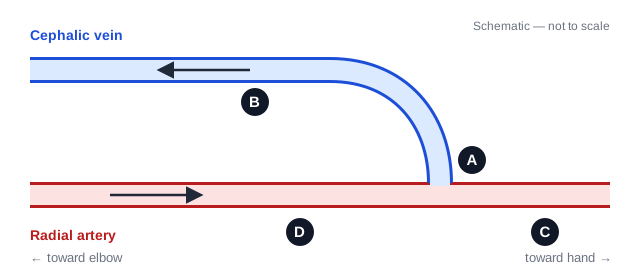
Schematic (original, not a sonogram): a radiocephalic arteriovenous fistula in the forearm.
Read diagram description
The radial artery runs left to right, from the elbow side to the hand side. A vein leaves the top of the artery near the right and runs up and then left toward the elbow. A: the point where the vein meets the artery. B: the vein, on the elbow side. C: the artery beyond the junction, toward the hand. D: the artery on the elbow side of the junction, where an arrow shows flow moving toward the junction. An arrow in the vein shows flow moving toward the elbow.
Terms: An anastomosis is a connection between vessels; inflow brings blood into the access and outflow carries it away.
Show answer and explanation
Answer: D. Region D
The inflow (feeding) artery is the artery upstream of the anastomosis. In this radiocephalic fistula, that's the radial segment on the elbow side of the connection — D, where the arrow shows flow heading into the junction. A is the arteriovenous anastomosis itself, B is the outflow (draining) cephalic vein, and C is the radial artery continuing toward the hand past the anastomosis. A complete access duplex looks at all three parts: inflow artery, anastomosis, and outflow vein.
Review topic: AV access anatomy
Domain: Surgically Altered Anatomy and Pathology
Question ID: RVT-PT-33
Sources:
- ACR–AIUM–SRU Practice Parameter for the Performance of Vascular Ultrasound Examinations for Postoperative Assessment of Hemodialysis Access — Specification of the Examination: forearm AVFs; examination of inflow artery, anastomosis, and venous outflow; Doppler angle alignment.
Question 34
Surveillance of an infrainguinal vein bypass graft shows a focal stenosis with a PSV of 350 cm/s and a velocity ratio of 3.8 compared with a nearby nonstenotic proximal graft segment. How is this usually classified?
Show answer and explanation
Answer: B. High-risk stenosis that warrants consideration of intervention
A PSV above 300 cm/s with a velocity ratio above 3.5 identifies a high-risk graft stenosis linked to graft failure. Measurable flow excludes complete occlusion at the sampled site, and nothing here describes arterialized venous flow.
Review topic: Vein graft surveillance
Domain: Surgically Altered Anatomy and Pathology
Question ID: RVT-PT-34
Sources:
-
SVS Guidelines Bundle: Follow-Up After Vascular Surgery Arterial Procedures — Page 6, Table 2: DUS Velocity and ABI Threshold Criteria for Stratification of Risk for Thrombosis of Infrainguinal Vein Grafts.
-
Tinder CN, Bandyk DF. Detection of imminent vein graft occlusion: what is the optimal surveillance program? — Abstract, critical-stenosis criteria and consideration of intervention.
Question 35
Why shouldn't native-artery velocity criteria be applied directly to a carotid artery after stenting?
Show answer and explanation
Answer: A. Stented segments tend to show higher velocities than native arteries with the same narrowing, so native criteria overestimate stenosis
A stent makes the artery stiffer and less compliant, so velocities run higher than in a native artery with the same degree of narrowing. Native criteria therefore overestimate in-stent stenosis, and modified, stent-specific thresholds are used. Stents are well seen on ultrasound; stenting does not inherently reverse flow or keep velocities below 50 cm/s.
Review topic: Carotid stent velocities
Domain: Surgically Altered Anatomy and Pathology
Question ID: RVT-PT-35
Sources:
-
SVS Practice Guidelines: Follow-Up After Vascular Surgery Arterial Procedures, overview slides — PDF page 11, carotid artery stenting recommendation 3.
-
Szegedi I et al. Role of carotid duplex in the assessment of carotid artery restenosis after endarterectomy or stenting — Discussion: Duplex ultrasound criteria for restenosis after CAS compared to stenosis in native ICA.
Question 36
After a liver transplant, the intrahepatic hepatic artery shows a tardus-parvus waveform with a resistive index of 0.45 and an acceleration time of 0.12 seconds. What should be suspected?
Terms: RI = resistive index.
Show answer and explanation
Answer: C. Hepatic artery stenosis upstream of the sampled site
A tardus-parvus waveform — RI below 0.5 with an acceleration time above 0.08 s — downstream of the anastomosis suggests upstream hepatic artery stenosis. It is a reason for further evaluation, not a diagnosis by itself; this waveform can give false-positive results. A normal hepatic artery has a brisk upstroke. The hepatic and portal veins aren't the vessel sampled.
Review topic: Liver transplant hepatic artery
Domain: Surgically Altered Anatomy and Pathology
Question ID: RVT-PT-36
Sources:
- Tardus parvus waveforms in Doppler ultrasonography for hepatic artery stenosis after liver transplantation: can a new cut-off value guide the next step? — Introduction; Materials and methods—Study population and Doppler ultrasonography; Results—traditional TPW; Discussion.
Question 37
Systolic pressures: right brachial 150 mmHg, left brachial 136 mmHg; right posterior tibial 108 mmHg, right dorsalis pedis 96 mmHg. What is the right ABI, and how is it classified?
Terms: ABI = ankle-brachial index; PAD = peripheral artery disease.
Show answer and explanation
Answer: C. 0.72; abnormal
ABI = the higher ankle pressure in that leg ÷ the higher of the two brachial pressures = 108 ÷ 150 = 0.72. An ABI of 0.90 or less is abnormal and consistent with PAD. A used the lower ankle pressure (96 ÷ 150); B used the lower arm pressure (108 ÷ 136); D inverted the fraction.
Review topic: ABI calculation
Domain: Physiologic Exams
Question ID: RVT-PT-37
Sources:
-
American Heart Association: A Clinical Assessment of Peripheral Artery Disease for Those on the Primary Care Spectrum — PDF page 31 / slide 31: STEP #3—The Resting ABI.
-
2024 ACC/AHA multisociety guideline for lower extremity PAD: official slide set — PDF page 38, Resting ABI recommendation 2; PDF page 40, Exercise ABI recommendation 6; PDF page 41, segmental pressures recommendation 8; PDF pages 16 and 28, claudication definition.
Question 38
A patient's resting ABI is 0.95. How is this classified under the current ACC/AHA multisociety PAD guideline?
Show answer and explanation
Answer: D. Borderline
The categories are: 0.90 or less abnormal, 0.91–0.99 borderline, 1.00–1.40 normal, and above 1.40 noncompressible. In a patient with exertional leg symptoms, a borderline result is a reason for further testing such as an exercise ABI.
Review topic: ABI categories
Domain: Physiologic Exams
Question ID: RVT-PT-38
Sources:
- 2024 ACC/AHA multisociety guideline for lower extremity PAD: official slide set — PDF page 38, Resting ABI recommendation 2; PDF page 40, Exercise ABI recommendation 6; PDF page 41, segmental pressures recommendation 8; PDF pages 16 and 28, claudication definition.
Question 39
A patient has calf pain with walking, but the resting ABI is 0.98. Which test best evaluates for PAD?
Show answer and explanation
Answer: A. Exercise treadmill ABI, looking for a drop in ankle pressure after exercise
When symptoms suggest claudication but the resting ABI is normal or borderline, guidelines recommend an exercise treadmill ABI to unmask PAD. Venous and carotid duplex examine different vascular problems, and simply waiting to repeat the resting ABI does not evaluate the current exertional symptoms.
Review topic: Exercise ABI
Domain: Physiologic Exams
Question ID: RVT-PT-39
Sources:
- 2024 ACC/AHA multisociety guideline for lower extremity PAD: official slide set — PDF page 38, Resting ABI recommendation 2; PDF page 40, Exercise ABI recommendation 6; PDF page 41, segmental pressures recommendation 8; PDF pages 16 and 28, claudication definition.
Question 40
In segmental limb pressure testing, what pressure drop between adjacent levels in the same leg generally suggests significant intervening arterial disease?
Show answer and explanation
Answer: B. 20 mmHg or more
A drop of 20 mmHg or more between adjacent cuff levels in the same leg generally suggests significant disease between those levels. Interpret segmental pressures with the waveforms and the rest of the examination; a smaller difference alone does not exclude disease.
Review topic: Segmental pressures
Domain: Physiologic Exams
Question ID: RVT-PT-40
Sources:
-
Gerhard-Herman M et al. Guidelines for Noninvasive Vascular Laboratory Testing — PDF page 7 / journal page 961, Segmental Limb Pressure Interpretation.
-
2024 ACC/AHA multisociety guideline for lower extremity PAD: official slide set — PDF page 38, Resting ABI recommendation 2; PDF page 40, Exercise ABI recommendation 6; PDF page 41, segmental pressures recommendation 8; PDF pages 16 and 28, claudication definition.
Question 41
A blood pressure cuff whose bladder is too narrow for the limb tends to produce what error?
Show answer and explanation
Answer: D. Falsely high pressure
A cuff that's too small needs more pressure to compress the artery, so it artificially elevates the reading; a cuff that's too large gives an artificially low reading. That's why cuff size must match limb size at every level you measure.
Review topic: Cuff size error
Domain: Physiologic Exams
Question ID: RVT-PT-41
Sources:
- Muntner P et al. Measurement of Blood Pressure in Humans: A Scientific Statement From the American Heart Association — Cuff Size subsection, text preceding Table 2.
Question 42
In venous photoplethysmography (PPG), what does venous refill time measure?
Show answer and explanation
Answer: A. The time for skin blood volume to return to baseline after calf-muscle exercise empties the veins
After repeated calf contractions empty the leg veins, the PPG sensor tracks how long skin blood volume takes to return to baseline. Normally the veins refill slowly from arterial inflow (roughly 20 seconds or more); reflux refills them backward and shortens the time. B is pulse transit time, C is duplex reflux timing, and D isn't a measurement.
Review topic: Venous PPG refill
Domain: Physiologic Exams
Question ID: RVT-PT-42
Sources:
-
Saliba Júnior OA et al. Pre- and Postoperative Evaluation by Photoplethysmography in Patients Receiving Surgery for Lower-Limb Varicose Veins — §2 Materials and Methods: PPG protocol and baseline curve; Figure 1; §4 Discussion: normal VRT convention and differing study cutoffs.
-
Franceschi C. Who knows the rationale of the refilling time measured by plethysmography? — PDF page 1 / journal page 8, Pathophysiology significance of the refilling time; PDF page 2, Figure 1 caption.
Question 43
Which sensor is typically used to detect pulse return when measuring a toe pressure?
Show answer and explanation
Answer: C. A photoplethysmography (PPG) sensor on the toe tip
Toe pressures use a small digit cuff with a PPG sensor on the toe tip, which detects pulsatile changes in skin blood volume as the cuff deflates. The other options don't measure pressure at the digit.
Review topic: Toe pressure sensor
Domain: Physiologic Exams
Question ID: RVT-PT-43
Sources:
- Scanlon C et al. Interrater and intrarater reliability of photoplethysmography for measuring toe blood pressure and toe-brachial index in people with diabetes mellitus — Methods: toe pressure and TBI measurement procedure using PPG probe and small toe cuff.
Question 44
While assisting the proceduralist with ultrasound-guided thrombin injection of the femoral pseudoaneurysm shown, which labeled point is the appropriate needle-tip target?
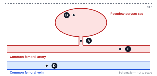
Schematic (original, not a sonogram): a femoral pseudoaneurysm with four candidate needle-tip points.
Read diagram description
Skin is at the top. A rounded pseudoaneurysm sac sits above the common femoral artery and connects to it through a narrow neck. The common femoral vein runs below the artery. A: inside the narrow neck. B: inside the sac, near its top edge and far from the neck. C: inside the common femoral artery. D: inside the common femoral vein.
Show answer and explanation
Answer: B. Point B
B is inside the sac and away from the neck. Keeping the tip away from the neck reduces the risk of thrombin entering the native artery through the neck (A) and causing distal thrombosis; it does not eliminate that risk. The artery (C) and the vein (D) are not injection targets for this procedure.
Review topic: Thrombin injection target
Domain: Ultrasound-guided Procedures/Intraoperative Assessment
Question ID: RVT-PT-44
Sources:
- Altuwaijri T et al. Ultrasound-guided thrombin injection versus ultrasound-guided compression repair in post-catheterization femoral artery pseudoaneurysm — Methods—Surgical technique: first paragraph for UGCR, third/fourth paragraphs for UGTI; Discussion: thromboembolic complications and native-artery occlusion.
Question 45
During ultrasound-guided compression repair of a femoral pseudoaneurysm, what is the goal of transducer pressure?
Show answer and explanation
Answer: D. Stop flow into the pseudoaneurysm at the neck while keeping flow in the native artery
Pressure over the neck stops flow into the sac so it can thrombose, while flow in the femoral artery is maintained. Occluding the native artery would risk limb ischemia.
Review topic: Compression repair
Domain: Ultrasound-guided Procedures/Intraoperative Assessment
Question ID: RVT-PT-45
Sources:
- Altuwaijri T et al. Ultrasound-guided thrombin injection versus ultrasound-guided compression repair in post-catheterization femoral artery pseudoaneurysm — Methods—Surgical technique: first paragraph for UGCR, third/fourth paragraphs for UGTI; Discussion: thromboembolic complications and native-artery occlusion.
Question 46
Before a planned great saphenous vein ablation, the proceduralist asks you to identify the saphenofemoral junction. Which labeled position marks that junction?
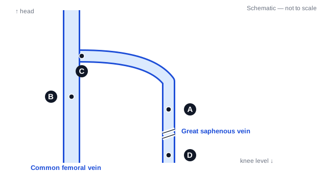
Schematic (original, not a sonogram): superficial and deep venous anatomy near the groin.
Read diagram description
The common femoral vein runs vertically on the left, head toward the top. The great saphenous vein runs down the leg on the right and curves up to meet the common femoral vein. A: in the great saphenous vein below the meeting point. B: inside the common femoral vein. C: at the point where the great saphenous vein meets the common femoral vein. D: far down the great saphenous vein at knee level.
Show answer and explanation
Answer: C. Position C
C marks the connection between the great saphenous vein and the common femoral vein: the saphenofemoral junction. Identifying this superficial-to-deep connection is part of mapping the anatomy before treatment. B is within the common femoral vein; A and D are along the great saphenous vein.
Review topic: Pre-ablation junction anatomy
Domain: Ultrasound-guided Procedures/Intraoperative Assessment
Question ID: RVT-PT-46
Sources:
-
AIUM Practice Parameter for the Performance of Peripheral Venous Ultrasound Examinations, 2024 Revision — Specification of the Examination §§1.1.2 and 1.2.1.2 (GSV/SFJ and CFV-GSV confluence); §1.3.3 (reverse Trendelenburg for venous distention).
-
SVS/AVF/AVLS clinical practice guidelines for varicose veins of the lower extremities, Part II — §1.4.3: complete venous reflux DUS including SFJ; §§11.1.1–11.1.4: early DUS after ablation; §11 introduction: ARTE terminology.
Question 47
A patient develops new leg pain and swelling after thermal ablation of the great saphenous vein. Why is an early duplex examination indicated?
Show answer and explanation
Answer: C. To assess for ablation-related thrombus extension (ARTE) or deep vein thrombosis
New symptoms after thermal ablation warrant early duplex to assess for ablation-related thrombus extension (ARTE) or deep vein thrombosis. ARTE includes the complication previously called endovenous heat-induced thrombosis (EHIT). The 2023 multisociety guideline recommends early duplex in symptomatic patients and advises it for high-risk patients; it recommends against routine early duplex solely to look for ARTE or DVT in average-risk, asymptomatic patients after thermal ablation. Vein graft measurement, confirming reopening, and an ABI do not address the suspected venous complication.
Review topic: Symptomatic post-ablation assessment
Domain: Ultrasound-guided Procedures/Intraoperative Assessment
Question ID: RVT-PT-47
Sources:
- SVS/AVF/AVLS clinical practice guidelines for varicose veins of the lower extremities, Part II — §1.4.3: complete venous reflux DUS including SFJ; §§11.1.1–11.1.4: early DUS after ablation; §11 introduction: ARTE terminology.
Question 48
An ICA spectral waveform looks high resistance with no diastolic flow, but color Doppler shows flow throughout the cycle. Which setting is most likely responsible?
Show answer and explanation
Answer: D. Wall filter set too high
A high wall filter strips out low-velocity signals near the baseline and can erase genuine diastolic flow, making a low-resistance vessel look high resistance. A too-low scale causes aliasing, not lost diastole. Center placement and a 50-degree angle are correct technique.
Review topic: Wall filter artifact
Domain: Quality Assurance, Safety, and Physical Principles
Question ID: RVT-PT-48
Sources:
- Bhide A et al. ISUOG Practice Guidelines (updated): use of Doppler velocimetry in obstetrics — Spectral pulsed-wave Doppler ultrasonography, wall-filter paragraph and Figure 4 caption: high filtering can obscure end-diastolic flow.
Question 49
Each panel shows the same vessel, flow direction, Doppler beam, and sample gate; only the angle-correction cursor changes. In which panel is the cursor aligned correctly for a velocity measurement?
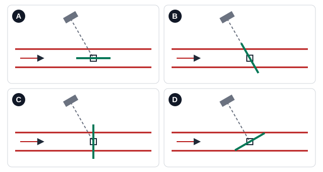
Schematic (original, not a sonogram): four placements of the angle-correction cursor.
Read diagram description
All four panels show the same straight horizontal vessel with flow from left to right, the same Doppler beam entering from above at 60 degrees to the flow, and a sample gate in the center of the vessel. Only the solid cursor line changes. A: the cursor lies parallel to the vessel walls and the flow. B: the cursor lies along the Doppler beam. C: the cursor is perpendicular to the flow. D: the cursor is tilted 30 degrees from the flow direction.
Show answer and explanation
Answer: A. Panel A
The angle-correction cursor belongs parallel to the direction of flow — here, parallel to the vessel walls — so the system uses the true angle between the beam and the flow (60° in every panel). B lines the cursor up with the beam, C sets it perpendicular to the flow, and D tilts it 30° off the flow. Each makes the machine calculate velocity with the wrong angle. Keep the beam-to-flow angle at 60° or less, because velocity errors grow quickly at larger angles.
Review topic: Angle-correction cursor
Domain: Quality Assurance, Safety, and Physical Principles
Question ID: RVT-PT-49
Sources:
- ACR–AIUM–SRU Practice Parameter for the Performance of Vascular Ultrasound Examinations for Postoperative Assessment of Hemodialysis Access — Specification of the Examination: forearm AVFs; examination of inflow artery, anastomosis, and venous outflow; Doppler angle alignment.
Question 50
An artery has a PSV of 90 cm/s, an end-diastolic velocity of 15 cm/s, and a time-averaged maximum velocity (TAMX) of 40 cm/s, measured from the spectral envelope over a cardiac cycle. What is the pulsatility index (Gosling)?
Terms: EDV = end-diastolic velocity; TAMX = time-averaged maximum velocity.
Show answer and explanation
Answer: B. 1.88
PI = (PSV − EDV) ÷ TAMX = (90 − 15) ÷ 40 = 1.875 ≈ 1.88. TAMX is the maximum-velocity envelope averaged over the cardiac cycle, not the intensity-weighted mean velocity. A is the resistive index, (90 − 15) ÷ 90. C divides PSV by TAMX; D divides PSV by EDV.
Review topic: Pulsatility index
Domain: Quality Assurance, Safety, and Physical Principles
Question ID: RVT-PT-50
Sources:
- Bhide A et al. ISUOG Practice Guidelines (updated): use of Doppler velocimetry in obstetrics — Which indices should be used?: PI = (S − D)/TAMX; distinction from intensity-weighted mean velocity; RI and S/D definitions.
Question 51
How is acceleration time measured on a spectral waveform?
Show answer and explanation
Answer: C. From the onset of systole to the first (early) systolic peak
Acceleration time runs from the start of the systolic upstroke to the early systolic peak. A prolonged acceleration time — a slow upstroke — suggests obstruction upstream of where you're sampling.
Review topic: Acceleration time
Domain: Quality Assurance, Safety, and Physical Principles
Question ID: RVT-PT-51
Sources:
- Trihan JE et al. Arterial Blood-Flow Acceleration Time on Doppler Ultrasound Waveforms: What Are We Talking About? — §2 Measurement Methods; §3 Proposal and Perspectives; Figure 1.
Question 52
While you scan the subclavian artery, a duplicate copy of the artery, with color flow, appears deep to the pleura. What artifact is this?
Show answer and explanation
Answer: B. Mirror image
The pleura–lung interface is a strong reflector that can produce a mirror-image duplicate of the subclavian artery, including its Doppler signal. Reverberation creates evenly spaced repeated lines; refraction creates edge shadows; aliasing is a Doppler wraparound.
Review topic: Mirror-image artifact
Domain: Quality Assurance, Safety, and Physical Principles
Question ID: RVT-PT-52
Sources:
- Rubin JM et al. Duplication images in vascular sonography — Abstract—five vascular duplication cases including subclavian arteries/vein, mirror-image multipath mechanism.
Question 53
A lab compares 100 arterial duplex studies with angiography. Angiography shows significant stenosis in 50; duplex correctly identified 45 of them. Of the 50 without stenosis, duplex called 4 positive. What is the duplex sensitivity?
Show answer and explanation
Answer: A. 90%
Sensitivity = true positives ÷ everyone with disease = 45 ÷ 50 = 90%. B is specificity (46 ÷ 50), C is positive predictive value (45 ÷ 49), and D is overall accuracy (91 ÷ 100). Correlation studies like this are how a lab checks its own criteria.
Review topic: Sensitivity and specificity
Domain: Quality Assurance, Safety, and Physical Principles
Question ID: RVT-PT-53
Sources:
- FDA. Statistical Guidance on Reporting Results from Studies Evaluating Diagnostic Tests — §7.1: estimates of sensitivity and specificity for a diagnostic test.
Question 54
During a lower-extremity arterial exam, the patient becomes short of breath and sweaty and reports chest pain. What should you do first?
Show answer and explanation
Answer: D. Stop the exam and get immediate help using the facility's emergency protocol
Possible acute cardiac or respiratory distress takes priority over the images. The sonographer monitors the patient's condition and activates the emergency protocol when needed.
Review topic: Patient monitoring
Domain: Quality Assurance, Safety, and Physical Principles
Question ID: RVT-PT-54
Sources:
- SDMS Scope of Practice and Clinical Standards for the Diagnostic Medical Sonographer — Clinical Standards §§1.4.5–1.4.6 and §1.5.4: monitor the patient and respond to emergencies.
Question 55
A requisition for a lower-extremity venous duplex lists "calf pain with walking, relieved by rest." What is the most appropriate action?
Show answer and explanation
Answer: B. Contact the ordering provider or supervising physician to confirm the appropriate exam
Exertional calf pain relieved by rest suggests arterial claudication. When the order doesn't fit the history, the sonographer clarifies it through facility policy with the supervising physician or referring provider — not by silently changing or cancelling it.
Review topic: Order appropriateness
Domain: Quality Assurance, Safety, and Physical Principles
Question ID: RVT-PT-55
Sources:
-
SDMS Scope of Practice and Clinical Standards for the Diagnostic Medical Sonographer — Clinical Standards §§1.1.1–1.1.2: verify the order and relevant history; clarify the examination.
-
2024 ACC/AHA multisociety guideline for lower extremity PAD: official slide set — PDF page 38, Resting ABI recommendation 2; PDF page 40, Exercise ABI recommendation 6; PDF page 41, segmental pressures recommendation 8; PDF pages 16 and 28, claudication definition.
Question 56
Which vascular exam most commonly calls for the patient to fast beforehand?
Show answer and explanation
Answer: A. Mesenteric artery duplex
Mesenteric duplex is done after fasting (commonly 6 hours or more) to reduce bowel gas and to remove the effect of digestion, which increases mesenteric flow. Carotid, upper-extremity venous, and ABI exams need no fasting.
Review topic: Patient preparation
Domain: Preparation, Documentation, and Communication
Question ID: RVT-PT-56
Sources:
- Society for Vascular Technology. Mesenteric Artery Duplex Ultrasound Examination guideline — PDF page 2: Patient preparation.
Question 57
Before starting an exam, how should you confirm the patient's identity?
Show answer and explanation
Answer: D. Use at least two patient identifiers, such as full name and date of birth
At least two person-specific identifiers, such as full name and date of birth, are the patient-safety standard, and a room number never counts as one. A yes/no question invites a wrong "yes," and someone else's confirmation doesn't replace your own check.
Review topic: Patient identification
Domain: Preparation, Documentation, and Communication
Question ID: RVT-PT-57
Sources:
- The Joint Commission. Two Patient Identifiers—Understanding the Requirements — FAQ response: two person-specific identifiers; room number is not an acceptable identifier.
Question 58
A clinically stable patient with heart failure has chronic orthopnea: lying flat causes breathlessness that resolves when the head is raised. For a lower-extremity venous duplex, what is the best positioning adaptation?
Show answer and explanation
Answer: C. Raise the head of the bed or use reverse Trendelenburg, and document the position used
Raising the head of the bed or using reverse Trendelenburg can improve tolerance in this stable patient. Reverse Trendelenburg also improves leg-vein distention for compression. Document the adaptation so the study can be interpreted and repeated. The exam should not be abandoned or narrowed solely because the patient cannot lie flat.
Review topic: Adapting the exam
Domain: Preparation, Documentation, and Communication
Question ID: RVT-PT-58
Sources:
-
AIUM Practice Parameter for the Performance of Peripheral Venous Ultrasound Examinations, 2024 Revision — Specification of the Examination §§1.1.2 and 1.2.1.2 (GSV/SFJ and CFV-GSV confluence); §1.3.3 (reverse Trendelenburg for venous distention).
-
SDMS Scope of Practice and Clinical Standards for the Diagnostic Medical Sonographer — Clinical Standards §1.3.1 and §1.4.2: adapt the examination to the patient; §§1.5.2 and 1.6.1–1.6.3: limitations and documentation.
Question 59
During a venous duplex, the patient asks, "Do I have a blood clot?" What is the best response?
Show answer and explanation
Answer: B. Explain that the interpreting physician will review the images and the results will come through the ordering provider, following facility policy
The sonographer refers specific diagnostic questions to the appropriate physician or healthcare provider and communicates results to the patient only as authorized by facility policy. A respectful explanation of the process avoids giving an unauthorized diagnosis, ignoring the patient, or falsely reassuring them.
Review topic: Patient questions and scope
Domain: Preparation, Documentation, and Communication
Question ID: RVT-PT-59
Sources:
- SDMS Scope of Practice and Clinical Standards for the Diagnostic Medical Sonographer — Clinical Standards §§1.2.3–1.2.4: patient questions and communication of results under facility authorization.
Question 60
At the end of a carotid duplex, which step belongs to completing the exam?
Show answer and explanation
Answer: D. Confirm the images are archived and document the preliminary findings and any technical limitations
Finishing the exam means making sure the images reached the archive and documenting preliminary findings and limitations for the interpreting physician. Deleting images loses the record, and the diagnosis comes from the interpreting physician.
Review topic: Completing documentation
Domain: Preparation, Documentation, and Communication
Question ID: RVT-PT-60
Sources:
- SDMS Scope of Practice and Clinical Standards for the Diagnostic Medical Sonographer — Clinical Standards §§1.5.2 and 1.6.1–1.6.3: documentation, technical limitations, preliminary findings, and archiving.
Use your result to choose what to review
Give yourself one point for each correct answer you chose before opening its explanation. For a complete-set score, unanswered questions and answers revealed before you chose an option receive zero points.
Practice percentage = correct answers ÷ 60 × 100. For example, 45 correct answers out of 60 is 75%.
Your result describes these 60 questions. It is not an ARDMS scaled score and cannot predict whether you will pass. Use missed and guessed questions to choose what to review next.
Start with two or three questions you missed or guessed. For each one, write down the clue that supports the best answer, why your choice did not fit, and the principle you need to review. Rework calculation questions from the inputs before looking at the formula again.
Find your review area
The set approximately follows the proportions in the official VT Content Outline v24.1, with whole-question rounding. It does not cover every detailed task in that outline. A domain with only four or five questions gives limited feedback, so use the individual explanations to guide your review.
| Review area | Official exam weight | Questions in this set |
|---|---|---|
| Normal Anatomy, Perfusion, and Function | 21% | 1–13 (13 questions) |
| Pathology, Perfusion, and Function | 32% | 14–32 (19 questions) |
| Surgically Altered Anatomy and Pathology | 6% | 33–36 (4 questions) |
| Physiologic Exams | 12% | 37–43 (7 questions) |
| Ultrasound-guided Procedures/Intraoperative Assessment | 7% | 44–47 (4 questions) |
| Quality Assurance, Safety, and Physical Principles | 14% | 48–55 (8 questions) |
| Preparation, Documentation, and Communication | 8% | 56–60 (5 questions) |
How this practice set compares with the VT exam
RVT is the credential; VT is the specialty examination. The official RVT pathway requires the applicable prerequisites and the SPI and VT examinations. This page practices vascular technology; the Sonography Principles and Instrumentation (SPI) exam is separate.
| Feature | This practice set | Official VT examination |
|---|---|---|
| Length | 60 questions | Approximately 170 questions |
| Time | Work at your own pace | Three hours; the official page separately lists a five-minute survey |
| Format | Single-best-answer questions, including four original schematics with A–D choices | Multiple-choice and hotspot questions |
| Result | Number correct and a practice percentage | A scaled score; 555 or higher passes on a 300–700 scale |
Sources: Official VT examination — “Question format & scoring” and official passing standard.
The diagrams are teaching schematics, not clinical sonograms or a reproduction of the testing interface. An official scaled score cannot be calculated from your percentage on this set. For the testing interface, Pearson’s ARDMS page provides a “Sample testing” tutorial link.
Sources and editorial information
By Castleport Test Prep Editorial Team.
Last verified: October 29, 2026. The exam format, scoring, linked VT outline, and the teaching principles cited with these 60 questions were checked against the identified sources. Source links and specific sections appear beside each explanation.
This resource was developed and checked with AI assistance. The checks covered sources, answer consistency, calculations, and editorial clarity; no credentialed clinical review is claimed. Read our methodology for how Castleport distinguishes these checks. These exercises support exam preparation and do not replace clinical training or supervised practice.
Castleport Test Prep is an independent exam prep publisher, not affiliated with, endorsed by, or approved by the American Registry for Diagnostic Medical Sonography (ARDMS). The questions and schematics are original, unofficial practice material, not actual ARDMS exam items. Exam and credential names identify their subjects; trademarks belong to their respective owners.